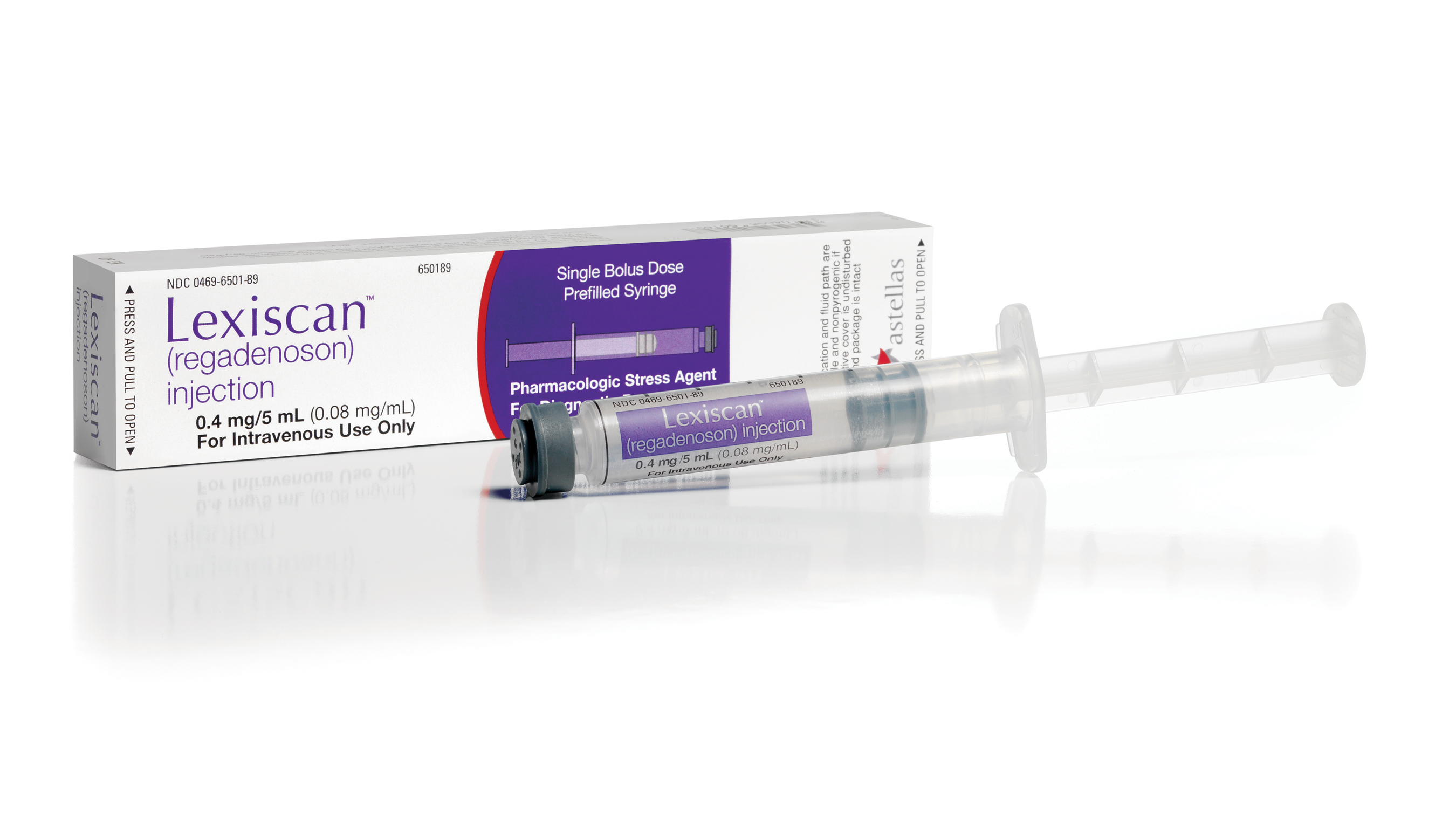
FDA最近通過一個新藥:lexiscan (regadenoson)
Lexiscan is a pharmacologic stress agent indicated for radionuclide myocardial perfusion imaging (MPI) in patients unable to undergo adequate exercise stress
Dosing Information
Drug Properties
A) Information
on specific products and dosage forms can be obtained by referring to the
Tradename List (Product Index)
B) Synonyms
Regadenoson
C) Physicochemical Properties
1) Molecular Weight
a) 408.37 (Prod Info LEXISCAN(TM) IV injection, 2008)
2) pH
a) 6.3 to 7.7 (Prod Info LEXISCAN(TM) IV injection, 2008)
Storage and Stability
A) Preparation
1) Intravenous route
a) Administer
regadenoson dose as a rapid intravenous injection, over approximately 10
seconds, into a peripheral vein using a 22 gauge or larger catheter or needle
(Prod Info LEXISCAN(TM) IV injection, 2008)
b) Immediately follow the
injection with a 5 milliliter saline flush (Prod Info LEXISCAN(TM) IV
injection, 2008).
c) Administer the radionuclide
myocardial perfusion imaging agent 10 to 20 seconds after the saline flush. The
radionuclide may be administered into the same catheter used to administer
regadenoson (Prod Info LEXISCAN(TM) IV injection, 2008).
B) Intravenous route
1) Solution
a) Store regadenoson solution at controlled room temperature, 25 degrees Celsius (77 degrees Fahrenheit); excursions permitted to 15 to 30 degrees Celsius (59 to 86 degrees Fahrenheit) (Prod Info LEXISCAN(TM) IV injection, 2008).
Adult Dosage
Normal Dosage
Intravenous route
Radionuclide myocardial perfusion study, Pharmacologic Stress
a) The recommended dose is 0.4 milligrams administered as a rapid (approximately 10 seconds) intravenous injection. Following an immediate 5 milliliter saline flush, administer the radionuclide myocardial perfusion imaging agent 10 to 20 seconds later (Prod Info LEXISCAN(TM) IV injection, 2008).
Dosage in Renal Failure
A) In studies that included patients with varying degrees of renal function (n=18) and healthy subjects (n=6), the fraction of regadenoson excreted unchanged in the urine as well as renal clearance of regadenoson were decreased with increasing degree of renal impairment (mild, creatine clearance (CrCl) 50 milliliters/minute (mL/min) or greater; moderate, CrCl 30 to less than 50 mL/min; severe, CrCl less than 30 mL/min). This led to increased elimination half-lives and AUC values compared to healthy subjects (CrCl, 80 mL/min or higher). However, as the Cmax and Vd values were similar across the groups and as the plasma concentration-time profiles were not significantly affected during the early stages following dosing, dosage adjustments are not necessary in patients with renal impairment. Regadenoson has not been evaluated in endstage renal disease patients on dialysis (Prod Info LEXISCAN(TM) IV injection, 2008).
Dosage in Hepatic Insufficiency
A) Pharmacokinetics of regadenoson have not been evaluated in patients with hepatic impairments. However, as greater than 55% of the dose is excreted unchanged in the urine and as factors that decrease clearance are not expected to result in clinically meaningful changes following regadenoson dosing, dosage adjustments are not necessary in patients with hepatic impairment (Prod Info LEXISCAN(TM) IV injection, 2008).
Dosage in Geriatric Patients
A) As the pharmacokinetics of regadenoson were not significantly influenced by age in population analysis, no dose adjustments are needed in elderly patients (Prod Info LEXISCAN(TM) IV injection, 2008).
Pediatric Dosage
Normal Dosage
Intravenous route
1) The safety and efficacy of regadenoson have not been established in patients less than 18 years of age (Prod Info LEXISCAN(TM) IV injection, 2008).
Clinical Applications
Monitoring Parameters
A) Therapeutic
1) Physical Findings
a) Reliably access the extent of reversible perfusion abnormalities via radionucleotide Single Photon Emission Computed Tomography (SPECT)
B) Toxic
1) Electrocardiograms
should be performed to monitor complications such as new arrhythmias, ischemia,
and atrioventricular (AV) and sinoatrial (SA) nodal conduction abnormalities
(Prod Info LEXISCAN(TM) IV injection, 2008).
2) Assess potential blood
pressure changes (Prod Info LEXISCAN(TM) IV injection, 2008).
3) Observe for signs or
symptoms of bronchoconstriction or respiratory compromise (Prod Info
LEXISCAN(TM) IV injection, 2008).
Patient Instructions
A) Regadenoson (Injection)
Regadenoson
Used for nuclear heart stress tests when the patient cannot exercise.
When This Medicine Should Not Be Used:
You should not use this medicine if you have had an allergic reaction to
regadenoson, or if you have certain heart problems (such as heart block or a
sinus node disorder) and do not have a pacemaker that is working.
How to Use This Medicine:
Injectable
Your doctor will prescribe your exact dose and tell you how often it
should be given. This medicine is given through a needle placed in one of your
veins.
A nurse or other trained health professional will give you this medicine.
Drugs and Foods to Avoid:
Ask your doctor or pharmacist before using any other medicine, including
over-the-counter medicines, vitamins, and herbal products.
Make sure your doctor knows if you are using dipyridamole
(Persantine®) before you receive this medicine. You may need to stop using
dipyridamole for at least two days before the test.
Make sure your doctor knows if you are using theophylline (Theo-24®, Uniphyl®)
before you receive this medicine. You may need to stop using theophylline for
at least twelve hours before the test.
Do not take anything that contains caffeine for at least twelve hours before
you receive this medicine. This includes medicines, foods, and beverages with
caffeine, such as coffee, tea, and cola drinks.
Warnings While Using This Medicine:
Make sure your doctor knows if you are pregnant or breastfeeding, or
if you have low blood pressure, breathing problems, or certain lung diseases
(such as asthma or chronic obstructive pulmonary disease [COPD]).
Tell your doctor right away if you start having chest pain, trouble with
breathing, lightheadedness, or if you feel faint after this medicine is
injected. You may be having a serious side effect from this medicine.
Possible Side Effects While Using This Medicine:
Call your doctor right away if you notice any of these side effects:
Allergic reaction: Itching or hives, swelling in your face or hands,
swelling or tingling in your mouth or throat, chest tightness, trouble
breathing.
Chest pain or discomfort.
Fast, slow, or uneven heartbeat.
Lightheadedness, dizziness, or fainting.
Pain, itching, burning, swelling, or a lump under your skin where the needle is
placed.
Shortness of breath.
If you notice these less serious side effects, talk with your doctor:
Change in taste.
Headache.
Nausea or upset stomach.
Warmth or redness in your face, neck, arms, or upper chest.
If you notice other side effects that you think are caused by this medicine,
tell your doctor.
Place In Therapy
A) Regadenoson is indicated as a pharmacologic stress agents for radionuclide myocardial perfusion imaging (MPI) in patients unable to undergo adequate exercise stress. Efficacy and adverse reactions were similar to adenosine in 2 randomized, double-blind clinical trials that included adult patients (n=2105) with known or suspected coronary artery disease requiring pharmacological stress MPI. Study patients had a variety of cardiovascular histories including hypertension (81%); CABG, PTCA, or stenting (51%); angina (63%), and history of MI (41%) or arrhythmia (33%) (Prod Info LEXISCAN(TM) IV injection, 2008).
Mechanism of Action / Pharmacology
A) Mechanism of Action
1) Regadenoson,
an A(2A) adenosine receptor agonist, produces coronary vasodilation and increases
coronary blood flow. It also causes a rapid increase in coronary blood flow
that lasts for a short duration. Mean average peak velocity of coronary blood
flow is increased to greater than twice the baseline level by 30 seconds and
decreased to less than twice the baseline level within 10 minutes (Prod Info
LEXISCAN(TM) IV injection, 2008).
2) The affinity of regadenoson
for the A(2A) adenosine receptor is low (K(i) approximates 1.3 micromoles
(mcM)), with at least a 10-fold lower affinity for the A(1) adenosine receptor
(K(i) greater than 16.5 mcM). Its affinity for the A(2B) and A(3) adenosine
receptors, if any, is weak (Prod Info LEXISCAN(TM) IV injection, 2008).
Therapeutic Uses
Radionuclide myocardial perfusion study, Pharmacologic Stress
FDA Labeled Indication
1) Overview
FDA Approval: Adult, yes; Pediatric, no
Efficacy: Adult, Effective
Recommendation: Adult, Class IIb
Strength of Evidence: Adult, Category B
See Drug Consult reference:
RECOMMENDATION AND EVIDENCE RATINGS
2) Summary:
Indicated as a pharmacologic stress agents for radionuclide
myocardial perfusion imaging in patients unable to undergo adequate exercise
stress (Prod Info LEXISCAN(TM) IV injection, 2008)
Administration of regadenoson demonstrated similar efficacy as adenosine in
radionuclide myocardial perfusion imaging in 2 randomized, double-blind
clinical trials in adult patients with known or suspected coronary artery
disease (Prod Info LEXISCAN(TM) IV injection, 2008)
In a multicenter, double-blinded phase 3 trial (n=784) of patients undergoing
myocardial perfusion imaging (MPI) studies, the strength of agreement between
sequential adenosine-regadenoson images was not inferior to the strength of
agreement between 2 sequential adenosine images for detecting the presence and
extent of reversible defects (Iskandrian et al, 2007).
3) Adult:
a) In a
multicenter, double-blinded phase 3 trial (n=784) of patients undergoing
myocardial perfusion imaging (MPI) studies, the strength of agreement between
sequential adenosine-regadenoson images was not inferior to the strength of
agreement between 2 sequential adenosine images, showing that regadenoson
provides diagnostic MPI comparable to a standard adenosine infusion for
detecting the presence and extent of reversible defects. Patients underwent an
initial qualifying imaging study with adenosine followed by a subsequent
randomized study with either regadenoson (n=517) or adenosine (n=267).
Regadenoson was given as a rapid bolus (less than 10 seconds) of 400
micrograms, adenosine was infused intravenously for 6 minutes at 140
micrograms/kilogram/minute. Based on the extent of reversible defects, each
scan was classified into one of three categories by use of a 17-segment model:
0 to 1, 2 to 4, or 5 or more reversible defects. The primary outcome was to
show noninferiority by demonstrating that the difference in the strength of
agreement in detecting reversible defects, based on blinded reading, between
sequential adenosine-regadenoson images and adenosine-adenosine images, lay above
a prespecified noninferiority margin (no reduction or a reduction of less than
13.33%; null hypothesis of inferiority is rejected when the lower limit is
above -13.33%). Average agreement rates, based on the median of 3 independent
blinded readers, between adenosine-adenosine images (n=259) and
regadenoson-adenosine images (n=499), were 0.64 +/- 0.04 and 0.63 +/- 0.03,
respectively. The difference was 1% (95% confidence interval (CI), -11.2% to
8.7%) with the lower limit of the CI being above the prespecified
noninferiority margin of -13.33%, demonstrating that regadenoson provides
diagnostic imaging comparable to adenosine for detecting the presence and
extent of reversible defects. Based on the presence or absence of reversible
defects, agreement rates between regadenoson-adenosine and adenosine-adenosine
were 82% versus 82% and 70% versus 69%, respectively (mean, 76% vs 76%). There
were no serious adverse events reported in the study groups and incidences of
adverse events were similar or fewer with regadenoson compared with adenosine.
The peak increase in heart rate was greater for regadenoson than adenosine, but
blood pressure responses were similar between the 2 groups. A tolerability
comparison, referred to as the summed symptom score (score based on presence or
severity of flushing, chest pain, and dyspnea), was significantly lower for
regadenoson compared to adenosine (p=0.013). Second-degree AV block was
reported in 3 patients with adenosine and none with regadenoson (p=0.043)
(Iskandrian et al, 2007).
b) In 2 randomized,
double-blind, comparative clinical trials (studies 1 and 2), administration of
regadenoson demonstrated similar efficacy as adenosine in radionuclide
myocardial perfusion imaging (MPI). Both studies included 2015 patients with
known or suspected coronary artery disease who were indicated for
pharmacological stress MPI. Patients (median age, 66 years; range, 26 to 93
years) received an initial stress scan using a 6-minute infusion of 0.14 milligrams/kilogram/minute
of adenoscan, without exercise, along with a radionuclide gates SPECT imaging
protocol. Following the initial scan, patients were randomized to receive
either regadenoson (n=1337) or adenosine (n=678) and a second scan was
conducted using the same radionuclide imaging protocol as the first scan. The
median time between the first and second scans was 7 days (range, 1 to 104
days). At baseline, the most common cardiovascular histories among study
patients were hypertension (81%); CABG, PTCA, or stenting (51%); angina (63%),
and history of MI (41%) or arrhythmia (33%). Cardioactive medications taken on
the day of the scan included beta blockers (18%), calcium channel blockers
(9%), and nitrates (6%). The scans were evaluated using the 17-segment model,
where the number of segments displaying a reversible perfusion defect was
calculated following the first and second scan. Agreement rates for the images
obtained after the first and second scans were compared and were based on the
frequency of patients assigned to each category within the 17-segment model
(0-1, 2-4, 5-17 segments). Based on pooled study data, 68% of patients had 0-1
segments displaying reversible defects on the initial scan; 24% had 2-4
segments, and 9% had 5 or more segments. In study 1, the mean +/- standard
error (SE) agreement rates after the second scan were 61% +/- 3% and 62% +/- 2%
in the adenosine and regadenoson groups, respectively (difference +/- SE, 1%
+/- 4%; 95% confidence interval (CI), -7.5% to 9.2%). In study 2, the mean +/-
SE agreement rates after the second scan were 64% +/- 4% and 63% +/- 3% in the
adenosine and regadenoson groups, respectively (difference +/- SE, -1% +/- 5%;
95% CI, -11.2% +/- 8.7%). Adverse reactions were similar in both groups.
Commonly reported adverse events in the regadenoson group that occurred at a
higher frequency than the adenosine group included dyspnea (28% vs 26%) and
headache (26% vs 17%). Among evaluable patients, rhythm or conduction
abnormalities occurred in 26% and 30% of patients in the regadenoson and
adenosine groups, respectively, first-degree atrioventricular block occurred in
3% and 7% of patients, respectively, and second-degree atrioventricular block
occurred in 0.1% and 1% of patients, respectively (Prod Info LEXISCAN(TM) IV
injection, 2008).
Comparative Efficacy / Evaluation With Other Therapies
Adenosine
Radionuclide myocardial perfusion study, Pharmacologic Stress
a) In a multicenter, double-blinded phase 3 trial (n=784) of patients undergoing myocardial perfusion imaging (MPI) studies, the strength of agreement between sequential adenosine-regadenoson images was not inferior to the strength of agreement between 2 sequential adenosine images, showing that regadenoson provides diagnostic MPI comparable to a standard adenosine infusion for detecting the presence and extent of reversible defects. Patients underwent an initial qualifying imaging study with adenosine followed by a subsequent randomized study with either regadenoson (n=517) or adenosine (n=267). Regadenoson was given as a rapid bolus (less than 10 seconds) of 400 micrograms, adenosine was infused intravenously for 6 minutes at 140 micrograms/kilogram/minute. Based on the extent of reversible defects, each scan was classified into one of three categories by use of a 17-segment model: 0 to 1, 2 to 4, or 5 or more reversible defects. The primary outcome was to show noninferiority by demonstrating that the difference in the strength of agreement in detecting reversible defects, based on blinded reading, between sequential adenosine-regadenoson images and adenosine-adenosine images, lay above a prespecified noninferiority margin (no reduction or a reduction of less than 13.33%; null hypothesis of inferiority is rejected when the lower limit is above -13.33%). Average agreement rates, based on the median of 3 independent blinded readers, between adenosine-adenosine images (n=259) and regadenoson-adenosine images (n=499), were 0.64 +/- 0.04 and 0.63 +/- 0.03, respectively. The difference was 1% (95% confidence interval (CI), -11.2% to 8.7%) with the lower limit of the CI being above the prespecified noninferiority margin of -13.33%, demonstrating that regadenoson provides diagnostic imaging comparable to adenosine for detecting the presence and extent of reversible defects. Based on the presence or absence of reversible defects, agreement rates between regadenoson-adenosine and adenosine-adenosine were 82% versus 82% and 70% versus 69%, respectively (mean, 76% vs 76%). There were no serious adverse events reported in the study groups and incidences of adverse events were similar or fewer with regadenoson compared with adenosine. The peak increase in heart rate was greater for regadenoson than adenosine, but blood pressure responses were similar between the 2 groups. A tolerability comparison, referred to as the summed symptom score (score based on presence or severity of flushing, chest pain, and dyspnea), was significantly lower for regadenoson compared to adenosine (p=0.013). Second-degree AV block was reported in 3 patients with adenosine and none with regadenoson (p=0.043) (Iskandrian et al, 2007).


 留言列表
留言列表
 線上藥物查詢