西弗(Sievert,Sv)是輻射劑量單位,
人體所受的輻射劑量是以輻射場強度與暴露時間相乘計算,常見單位有「微西弗╱時」及「毫西弗╱年」兩種。1毫西弗(millisievert,mSv)等於1000微西弗(microsievert,μSv)。
原子能委員會輻射偵測中心主任黃景鐘指,普通人1年可承受輻射量國際標準上限為1毫西弗,不過此標準十分嚴格,如果超過,不會有立即危險,但若在數天的短時間內受到4000毫西弗污染,就會有白血球減少等立即危險。
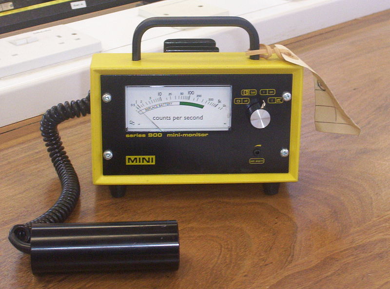
蓋格計數器(Geiger counter)又叫蓋格-米勒計數器(Geiger-Müller counter),是一種用於探測電離輻射的粒子探測器,通常用於探測α粒子和β粒子,也有些型號蓋格計數器可以探測γ射線及X射線。

構造及原理
蓋格計數器是根據射線對氣體的電離性質設計成的。其探測器(稱「蓋格管」)的通常結構是在一根兩端用絕緣物質密閉的金屬管內充入稀薄氣體(通常是摻加了鹵素的稀有氣體,如氦、氖、氬等),在沿管的軸線上安裝有一根金屬絲電極,並在金屬管壁和金屬絲電極之間加上略低於管內氣體擊穿電壓的電壓。這樣在通常狀態下,管內氣體不放電;而當有高速粒子射入管內時,粒子的能量使管內氣體電離導電,在絲極與管壁之間產生迅速的氣體放電現象,從而輸出一個脈衝電流信號。通過適當地選擇加在絲極與管壁之間的電壓,就可以對被探測粒子的最低能量,從而對其種類加以甄選。
蓋格計數器也可以用於探測γ射線,但由於蓋格管中的氣體密度通常較小,高能γ射線往往在未被探測到時就已經射出了蓋格管,因此其對高能γ射線的探測靈敏度較低。在這種情況下,碘化鈉閃爍計數器則有更好的表現。
歷史
蓋格計數器最初是在1908年由德國物理學家漢斯·蓋格和著名的英國物理學家盧瑟福在α粒子散射實驗中,為了探測α粒子而設計的。後來在1928年,蓋格又和他的學生米勒(Walther Müller)對其進行了改進,使其可以用於探測所有的電離輻射。
1947年,美國人Sidney H. Liebson在其博士學位研究中又對蓋格計數器做了進一步的改進[2],使得蓋格管使用較低的工作電壓,並且顯著延長了其使用壽命。這種改進也被稱為「鹵素計數器」。
蓋格計數器因為其造價低廉、使用方便、探測範圍廣泛,至今仍然被普遍地使用於核物理學、醫學、粒子物理學及工業領域。
Up to date的輻射劑量介紹:
UNITS OF RADIATION DOSE — The amount of radiation (ie, radiation dose) absorbed by the patient's tissues is highly predictive of its biological effects. Such doses are defined as the amount of energy of ionizing radiation deposited per unit of tissue mass at a specific point .
It is important to distinguish ionizing radiation (eg, x-rays, gamma rays, proton beams used for radiation therapy) from non-ionizing radiation (eg, microwaves, radio waves, infrared light). Nonionizing radiation generally causes damage through direct or indirect transfer of thermal (heat) energy; sunburn and microwave heating are classic examples of such exposures. On the other hand, ionizing radiation acts at the cellular level and has the potential to cause structural and chemical damage to vital targets such as nucleic acids and proteins.
The terms most often used for quantifying radioactivity, radiation dose, and radiation injury are described in the table (table 1). Those that will be employed in this review are defined below.
Absorbed Dose — The rad (radiation absorbed dose) is the traditional unit of absorbed dose, and is defined as the transfer of 100 ergs per gram of tissue. The rad has been superseded in the SI (Système International) by the Gray (Gy). One Gy, the unit most commonly used to measure radiation therapy dose, is equivalent to 100 rad (1 joule/kilogram), while one cGy is equivalent to 1 rad or 1000 mrad.
Dose Equivalent — The rem (Roentgen equivalent in man) is a unit for the dose equivalent and represents the product of the absorbed dose (in rads) and weighting factors that take into account the differential sensitivity among tissues as well as the biological effectiveness ("quality factor") of various sources of ionizing radiation . The rem has been superseded in the SI by the Sievert (Sv). One Sv is equivalent to 100 rem.
For most therapeutic radiation exposures (eg, x-rays, gamma rays) the Sievert and Gray are approximately equal. However, when there is exposure to highly ionizing particles (eg, neutrons, alpha particles) the radiation dose equivalent reflects resulting tissue damage better than the absorbed dose. As an example, the quality factor for x-rays, gamma rays, and beta particles is 1, while that for alpha particles is 20, and can range from 4 to 22 for neutrons, depending on neutron energy.
Dose rate — The "dose rate" refers to the amount of radiation delivered per unit of time and is most often measured in rads/hour or Gy/hour. Geiger counters typically provide an estimate of dose rate that may, in turn, be used to estimate the degree of hazard in a particular environment (eg, accident scene, patient clothing or bodily wastes). Common, hand-held Geiger counters are suitable for monitoring radioactive sources emitting gamma rays, but more specialized equipment is required for sources emitting certain low energy beta particles, neutrons, and alpha particles.
For cases involving exposure to x-rays, gamma rays and beta particles, a reduction in the radiation dose rate results in a decreased radiation response. For example, it is believed that the carcinogenic effect of these radiations delivered at a lower dose rate is less than that of the same total dose delivered at a higher dose rate. Similarly, a dose of 1 Gy delivered over 1 minute might cause signs and symptoms of acute radiation injury, whereas the same dose delivered over 100 days would not .
Examples of possible radiation exposures — The average annual dose to persons residing in the United States is approximately 3.6 mSv (360 mrem) . The majority of this dose (55 percent) is due to exposure to radon daughter products from the earth and construction materials, with man-made sources of radiation (eg, medical imaging studies), cosmic radiation, and natural radiation from endogenous sources (eg, the naturally occurring radioactive isotope of potassium, potassium-40) contributing the majority of the rest.
Examples of the ranges of exposures that might be seen following medical imaging procedures include the following :
- A standard chest x-ray delivers a dose of 6 to 11 mrem (0.06 to 0.11 mSv, 0.06 to 0.11 mGy).
- Interventional cardiologists working in a high-volume catheterization laboratory may have collar badge exposures exceeding 600 mrem (6 mSV) per year .
- A barium enema with 10 spot images delivers a dose of approximately 0.7 rem (700 mrem, 7 mSv, 7 mGy). Similar doses (7 to 8 mSv) are delivered from a CT scan of the chest or a PET scan, while a combined PET/CT scan is estimated to deliver a dose of 25 mSv [8].
The biologic effect of radiation doses higher than those achieved after routine medical imaging procedures are outlined below:
- The lowest radiation dose resulting in an observable effect on bone marrow depression in man, with a resultant decrease in blood cell counts, is in the range of 10 to 50 rem (100 to 500 mSv, 0.1 to 0.5 Gy).
- The lowest total body dose at which the first deaths may be seen following exposure to ionizing radiation is in the range of 1.0 to 2.0 Gy. Depending upon the type of support given, 50 percent of people exposed to a dose of 3 to 4 Gy will be expected to die of radiation-induced injury.
- There is virtually no chance of survival following a total body exposure in excess of 10 to 12 Gy.
The risk of radiation-induced carcinogenesis and other adverse health effects from medical imaging is controversial and is discussed separately.
關於輻射計量的名詞:
Terms employed for measurement of ionizing radiation
| Curie (Ci) |
| The amount of material whose atoms are undergoing a certain number of radiaoactive decays per unit time. One Curie, which was originally set as the number of atomic disintegrations per second in 1 gram of radium, is equal to the amount of radiactive material undergoing 3.7 x 10(10) disintegrations per second. This unit has been superceded by the Becquerel (Bq). One Becquerel is equal to the amount of material which is undergoing 1 disintegration/second. |
| Roentgen (R) |
| The amount of x-ray or gamma ray radiation which produces a certain number ofionizations (ion pairs) in a fixed quantity of air. One Roentgen is the quantity of x- or gamma-radiation which produces, in 0.001293 grams of air, ions carrying one electrostatic unit of electrical charge. |
| Roentgen equivalent in man (rem) |
| One rem is the amount of ionizing radiation of any type which produces in man the same biologic effect as one Roentgen of x- or gamma-rays. It is equal to the absorbed dose, measured in rads, multiplied by the relative biologic effectiveness (rbe) of the radiation in question. As examples, the rbe of x- and gamma rays is 1; the rbe for alpha particles and neutrons is 20. This unit has been superceded by the Sievert (Sv). One Sievert is equal to 100 rem. |
| Radiation absorbed dose (rad) |
| One rad is equal to the absorption of 100 ergs of ionizing radiation per gram of tissue. This unit has been superceded by the Grey (gy). One Grey is equal to 100 rad. |
致死的輻射劑量
Lethal dose of radiation — Estimation of the dose associated with death in 50 percent of those similarly exposed (ie, the LD 50) have been made in various scenarios. As an example, virtually all survivors of the explosion of a nuclear device at Hiroshima had estimated exposures of less than 3 Gy . Depending on the incident, estimates for the LD 50 have ranged from 1.4 Gy among atomic bomb survivors in Japan to 4.5 Gy following uniform total-body exposure to external photons . Several factors determine the lethality of ionizing radiation. These include:
- Dose rate — doses received over a shorter period of time cause more damage.
- Distance from the source — For point sources of radiation, the dose rate decreases as the square of the distance from the source (inverse square law).
- Shielding — Shielding can reduce exposure, depending upon the type of radiation and the material used [12]. As examples, alpha particles can be stopped by a sheet of paper or a layer of skin, beta particles by a layer of clothing or less than one inch of a substance such as plastic, and gamma rays by inches to feet of concrete or less than one inch of lead.
- Available medical therapy — The availability of supportive therapy (eg, antibiotics, transfusion, use of cytokines, hematopoietic cell transplantation) is critical for those exposed to moderately high doses of radiation. These subjects are discussed separately.
Based upon an analysis of all available data, the LD 50 at 60 days (LD 50/60) for humans has been estimated to be approximately 3.5 to 4.0 Gy in persons managed without supportive care, 4.5 to 7 Gy when antibiotics and transfusion support are provided, and potentially as high as 7 to 9 Gy in patients with rapid access to intensive care units, reverse isolation, and hematopoietic cell transplantation


 留言列表
留言列表
 線上藥物查詢