


BRB,Blood retinal barrier,血液-視網膜 障壁。
簡單的說就是在視網膜血管和一般血管中間的一個分隔區。
你可能會對於BBB(Blood brain barrier)比較熟悉,其實人體中存在著很多這樣的屏障
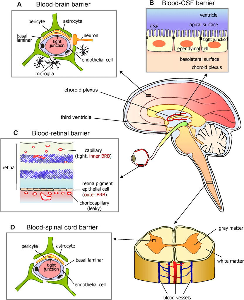
blood–testis barrier (BTB)

這樣的屏障多半都是靠著junction來進行篩選隔離的效果

在顯微鏡底下可以看到是這樣

Fenestrations
Cellular transport of material across the barrier can occur by two pathways: transcellular flux that is transport across the cell, or paracellular flux, transport between the cells.
Transcellular flux is transport through the cell that may be passive diffusion, facilitated, or by active transport mechanisms.
Fenestrations are locations in capillaries where the endothelial cell depth is reduced, and the thinning of the capillary wall facilitates transcellular transport of materi- als and cells out of the capillaries.
Fenestrations are seen in highly permeable tissues such as the glomerular capillary wall and choroid capillary plexus .
Endothelial cells in the BBB lack fenestrations contributing to their ability to maintain a barrier.
This loss of endothelial cell fenestrations is one of the initial developmental steps of BBB formation .
Although fenestrations have not been fully explored in the BRB, the analogous function and conservation of properties of the BRB and BBB suggest a similar paucity of fenestrations.
Endocytosis and Facilitated Diffusion
Cells continuously take up and sample extracellular material in a process called pinocytosis .
Membrane invaginations across the cell surface pinch off forming vesicles that move to the cell interior.
These vesicles can move internally to be degraded or through the cell to be released at the basal-lateral membrane leading to non-specific transport of materials across the cell.
Pinocytotic vesicles are selectively decreased in endothelial cells located in the BBB . In order to maintain transport of necessary substrates for neuronal function, specific receptor-facilitated transport mechanisms are used to move materials across the BBB.
Receptor mediated endocytosis may occur through clathrin or caveolin mediated endocytosis, which is ATP dependent.
A receptor binds to its specific substrate concentrating the substrate within the invagination to reduce transport of non-specific materials .
The transferrin receptor and the transport of iron across the BBB is an important example of receptor mediated transport across vascular endo- thelium .
Vascular endothelial cells of the BBB have increased mitochondria content, which gener- ates a cell’s energy supply of ATP.
Elevated mit- ochondria levels may be necessary to supply numer- ous receptor-mediated transport mechanisms.
Channel-facilitated transport is a mechanism for the diffusion of specific substrates across the BBB.
This is mediated by transmembrane proteins located in the cell surface that allow for the flux of specific materials across the cell membrane.
The GLUT-1 glucose transporter is highly and selectively ex pressed in vascular endothelial cells of the BBB and supplies the neuronal tissue with necessary glucose.
Together these mechanisms provide the necessary substrates for neuronal function.
Water Transport
Facilitated transport of water out of the neuronal extracellular space is essential to maintain retinal function.
RPE cells regulate water content and lactic acid removal generated by high metabolic rates in the retina. RPE cells transport water out of the retina and into the choroid capillary plexus.
The force gen- erated by water flux helps to maintain retinal attachment.
Lactic acid and water transport across the RPE are closely linked. The movement of water out of the retina is accomplished through the use of a number of ion channels and a specific water channel, aquaporin-1.
Aquaporin-1 is a transmembrane protein found in water permeable tissues that forms a transcellular channel for water transport.
Water transport is dependent on the pumping of Cl– and K+ across the RPE basolateral membrane into the choroid capillary plexus.
The flow of these ions is driven by the apical Na+/K+ATPase pump that elevates internal K+ levels allowing the subsequent flux of Cl–.
In addition, lac- tic acid is taken up by the RPE from the neuronal extracellular space by the monocarboxylate transporter-1 and then transported from the RPE cell interior to the choroid capillary plexus by monocar- boxylate transporter-3 .
Increased acid- ification of the RPE cell due to lactic acid buildup activates the Cl– and K+ pumps on the apical membrane, with subsequent diffusion across the basolateral membrane that ultimately drives water flux through aquaporin channels.
The reuptake of Cl– from the choroid capillary plexus by HCO3–/Cl– exchange pumps is activated with extracellular acidification.
In summary, elevated retinal metabolic rates increase lactic acid production. The RPE responds to this elevated metabolism by increasing lactate transport into RPE cells, which increases water transport from the retina to the choroid capillary plexus allowing for compensatory removal of by-products from the retina.
This regulation of transcellular transport requires precise control of paracellular flux between the two compartments, which is achieved by well-developed tight junctions.
(資料來源:Retinal Vascular Disease ISBN-13: 978-3540295419)
BRB主要分成兩個部分inner BRB(內層BRB)還有outer BRB(外層BRB)


Inner blood–retinal barrier
內層BRB的主要結構就是內層血管內皮細胞,Müller cell,tight junction還有pericyte(外被細胞)。
Müller cell又稱為Neuroglial cell,分布在photoreceptor和ganglion cell的軸突之間(從retina最內層延伸至cone cell的outer segment,橫跨2~10層,process axon長),是一種astrocyte(星狀細胞)
The endothelium of intraretinal blood vessels is considered as the main component of the inner blood–retinal barrier (BRB) resembling the blood–brain barrier in that both separate blood from neural parenchyma. The presence of a complex network of tight junctions, the absence of fenestrations and a relative paucity of caveolae make up the tightness of the iBRB. Numerous transport systems account for the selectivity of the barrier,such as the transport system for glucose across the retinal capillary endothelial cells of the inner BRB, which is mediated by the sodium-independent glucose transporter GLUT1.
Pericytes
多出現在小血管周圍,為一種未分化的mesenchymall cell,受刺激可增值分化為血管壁的平滑肌細胞或結締組織的細胞。主要為包圍為血管內皮細胞的細胞,可以產生收縮功能
Both pericytes and smooth muscle cells provide structural support to the vasculature. The increased density of pericytes in the retinal microvasculature as compared to other organs has led to the presumption that pericytes play a role in the regulation of retinal perfusion by contraction and relaxation.
Circumstantial evidence for contractility of pericytes is the expression of a number of contractile proteins, in particular α-smooth muscle actin, desmin, and non-muscle myosin.
Moreover, pericytes express receptors for vasoactive substances, enabling them to respond by contraction or relaxation. Recent work suggests that pericytes, rather than endothelial cells, control capillary diameter, because diameter changes do not occur in pericyte-free regions. Thus, pericytes would play a major role in redirecting blood flow at the capillary level.
Glial cell(神經膠細胞)
In the retina, both astrocytes and Müller cells participate in vessel ensheathment, whereby astrocytes are confined to the optic nerve fiber and the ganglion cell layer.[91] Glial cells play an important role in vessel integrity and barrier properties, both via direct contact[92] and through release of humoral factors.
Glial cell line-derived neurotrophic factor (GDNF) has been recognized to enhance barrier tightness,whereas transforming growth factor-β (TGF-β) appears to decrease it.
Several other growth factors and cytokines, such as tumor necrosis factor-α (TNF-α), interleukin-6 (IL-6), and vascular endothelial growth factor (VEGF) are produced by retinal glial cells and affect the tightness of the iBRB. VEGF directly downregulates tight junctional proteins, evoking a decrease in transendothelial resistance。
Outer blood–retinal barrier
外層BRB主要結構就是視網膜色素內皮細胞,tight junction,fenestrated endothelium of the choriocapillaris 開孔型微血管的有孔內皮細胞,Bruch's membrane。
The outer blood–retina barrier (BRB) is composed of three structural entities, the fenestrated endothelium of the choriocapillaris, Bruch's membrane, and the retinal pigment epithelium (RPE).

Fenestrated endothelium
The fenestrations of the choriocapillaris are covered by a thin membrane with a central thickening.
They have high permeability, similar to that of an isoporous membrane with a pore diameter of 4 Å,which probably accounts for the maintenance of an adequate concentration of glucose and other nutrients at the RPE level.
Retinal pigment epithelium
色素上皮內側形成突起,與photoreceptors(感光接受器)的outer segment交錯連接,吸收感光接受器之間的光線,以避免光線反射,而增加視覺敏感度。
色素上皮與外側choroid layer連接緊密,與內側photoreceptors附著力較弱
The RPE cells are equipped with an elaborate transcellular pathway system and an apico-lateral seal formed by intercellular junctions, the adherens junction and the tight and gap junctions, as well as, in some species, desmosomes.
The RPE is considered as a relatively tight epithelium with a 10 times higher paracellular than transcellular resistance
Nutrients and vitamin A are transported in basolateral to apical direction from the blood to the photoreceptors. In the opposite direction, transcellular transport from the subretinal space towards the choriocapillary bed is required for removal of metabolites, water, and ions. The transepithelial ion transport is linked to the transport of lactic acid, the major metabolic end product of neuronal function.
Bruch's membrane
Bruch membrane是一種base membrane,為視網膜和脈絡膜之間的纖維膜。
Using electron microscopy, five distinct layers of Bruch's membrane have been defined: the two basal laminas of the RPE and the endothelium of the choriocapillaris, the inner and outer collagenous layers, and a central discontinuous elastic layer.
Bruch's membrane provides tensile strength and, thanks to the presence of proteoglycans, constitutes a reservoir of growth factors. Its overall negative charge is due to the high content of glycoconjugates and it is responsible for charge-selective restriction to the passage of ions and solutes.

(A) Electronic micrography of the outer blood–retinal barrier. The retinal pigment epithelium (RPE) encloses the extremities of the external segments of the photoreceptors (PR) by means of long apical digitations, which are indicated by the two opposed arrows. At the basal pole, the plasma membrane forms numerous invaginations, the extremities of which rest on the basal lamina and form a part of Bruch's membrane (BM). PH = phagosome resulting from the internalization and lysosomal digestion of external segments. (Bar: 0.5 µm).
(B) Detail of the fenestrated endothelium of the choriocapillaris. The fenestrations of the endothelium cell (EC) are indicated by arrowheads. The contrast of the membranes and basal lamina was increased by tannic acid. (Bar: 0.1 µm).
圖片來源:From Rungger-Brändle & Leuenberger 2008.
綜合上面的通道,可以得知以下這張圖


 留言列表
留言列表
 線上藥物查詢