Endometriosis is a relatively common disease, affecting up to 10% of women of reproductive age.1
Its incidence is as high as 50% among adolescents with pelvic pain.1,2 Clinical symptoms include dysmenorrhea, pelvic pain, and infertility.1,3,4 Endometriotic lesions are considered to be benign (nonmalignant or nonneoplastic), inflammatory, estrogen-dependent lesions that are characterized by the ectopic presence of normal-appearing, functional endometrial tissue composed of glands and stroma outside of the uterus.1,3 The disease is often associated with multiple lesions that can be distributed throughout the abdominal–pelvic peritoneum and visceral organs.
There are three anatomical subtypes of endometriosis: superficial peritoneal endometriosis, ovarian endometriosis, and deep infiltrating endometriosis. Deep infiltrating endometriosis is characterized by nodules that locally invade pelvic structures, producing symptoms such as painful intercourse (deep dyspareunia) and painful bowel movements (dyschezia).5 Progestin-based hormonal therapy and gonadotropin-releasing hormone analogues have become the standard treatments; however, many patients have unacceptable systemic adverse effects in association with either or both treatments.1,6 Moreover, not all women with endometriosis have a response to hormonal therapy, particularly those who have deeply infiltrating disease.7 Surgical resection is an option for women who do not have a response to hormonal therapy or for those who desire pregnancy, but complete excision of deep infiltrating nodules requires surgical expertise and is not without risk.4
Despite its benign clinical behavior and normal-appearing histologic features, endometriosis can recapitulate some features of malignant neoplasms, including local invasion and resistance to apoptosis. The etiologic factors underlying endometriosis are controversial, and the disorder has been proposed to originate from several different processes: the migration of endometrial fragments from the uterus through the fallopian tubes during retrograde menstruation, the dissemination of these fragments to the peritoneal cavity, and their implantation on the serosal surface; the dissemination of endometrial (progenitor) cells through the lymphatic or blood circulation; the development of endometrial tissue through metaplasia of coelomic epithelium (i.e., mesothelium that lines the surface of the peritoneal cavity and organs); or differentiation from bone marrow–derived stem cells or fetal remnants of müllerian cells shed into the peritoneal cavity during retrograde menstruation.3,8-12
Genomewide association studies have identified genetic markers that are potentially related to an increased risk of endometriosis.13 Endometriosis, particularly ovarian endometriosis, is widely accepted as the direct precursor of clear-cell and endometrioid ovarian carcinomas,14,15 so-called endometriosis-related ovarian neoplasms. However, the study of somatic mutations in endometriosis has been restricted largely to endometriosis with concurrent cancer. A small number of candidate-gene studies have examined benign (non–cancer-associated) ovarian endometriosis lesions; one study identified a KRAS mutation (p.G12C) in a single lesion,16 and another identified PTEN mutations in 7 of 34 lesions (21%).17 Immunohistochemical studies have also shown rare partial or complete immunohistochemical loss of ARID1A (as a proxy for ARID1A loss-of-function mutations) in lesions from ovarian and nonovarian endometriosis without concurrent cancer.18-20 All of the above studies have been hampered by low-resolution genomic methods, failure to isolate specific endometrial stromal or epithelial cells from within a dominantly fibrotic endometriosis nodule (e.g., by laser-capture microdissection), and lack of orthogonal validation. Thus, these studies have yielded ambiguous answers to the fundamental question: do benign endometriosis lesions harbor somatic mutations in cancer driver genes outside of the process of transformation to endometriosis-related ovarian neoplasms, and if so, what is their role in the pathology of these lesions?
In this study, we performed exomewide sequencing and targeted amplicon sequencing and used two digital genomic methods to determine whether benign, but invasive, deep infiltrating endometriosis lesions harbor somatic mutations, including those frequently detected in human cancers.
METHODS
Tissue Samples and Patient Selection
Tissue samples were obtained from three independent cohorts of patients with deep infiltrating endometriosis. All tissues were fixed archival specimens, either formalin-fixed and paraffin-embedded or Molecular-fixed (Sakura Finetek) and paraffin-embedded. Tissue samples from 17 patients were obtained from the Departments of Obstetrics and Gynecology and Pathology at Lenox Hill Hospital–Northwell Health (Hofstra University) in New York. Tissue samples from 7 patients were obtained from the Department of Pathology at Seirei Mikatahara Hospital in Hamamatsu, Japan. Tissue samples from 15 patients were obtained from the BC Women’s Centre for Pelvic Pain and Endometriosis in Vancouver, BC, Canada, and collected through the OVCARE Tissue Bank (part of the World Endometriosis Research Foundation Endometriosis Phenome and Biobanking Harmonisation Project).21 The institutional review boards at the respective hospitals approved all tissue collection. Inclusion criteria were pathologist-confirmed deep infiltrating endometriosis (lesions with a depth >5 mm, such as in the bowel or peritoneal wall) containing both epithelial and stromal components, the absence of cancer or dysplasia, and a lesion size sufficient for tissue coring, macrodissection, or laser-capture microdissection.
Exome Sequencing
Endometriotic lesions and corresponding normal tissues from the same blocks were cored (2 mm for smaller lesions and 3 mm for larger lesions) from formalin-fixed and paraffin-embedded blocks. Sections that were stained with hematoxylin and eosin were prepared before and after coring to confirm the precision of the sampling and to ensure that at least 60% of the core area contained lesional tissue (or that 100% contained normal tissue). Paired-end libraries were generated with the use of standard Illumina procedures that involved DNA from core endometriosis and normal (matched control) samples. Coding regions were captured with the Agilent SureSelect Enrichment System and sequenced on Illumina sequencers. Data were processed as described previously.22
Probabilistic Evaluation of Observed Driver Mutation Frequencies
The probability that by pure chance, 5 mutations of the 80 that were found would be among the 125 driver genes considered was P=0.001, calculated by the binomial test (Table S1 in Supplementary Appendix 2, available with the full text of this article at NEJM.org). Specifically, 1–pbinom (4, 80, 276,824/27,000,000)=0.001, in which the total number of positions sequenced is approximately 27 million bp (whole exome), and 276,824 is the estimated total number of positions, among all the driver genes considered, that may yield a driver mutation. The number of driver positions was derived by combining all positions found to be (somatically) mutated, across all cancer types, in the Cancer Genome Atlas database that are considered to be located within driver oncogenes. We conservatively assumed that only targeted positions in tumor suppressor genes were targets of driver mutations.
Targeted Sequencing
The TruSeq Amplicon Cancer Panel (Illumina) and a previously described TruSeq Custom Amplicon Panel23 were applied, followed by Illumina MiSeq sequencing (Table S5 in Supplementary Appendix 2). Variants were identified with the use of the MiSeq Reporter Somatic Variant Caller tool. Only variants absent from germline DNA and present in both libraries (technical replicates) from both panels (i.e., quadruplicate libraries) were validated orthogonally by a droplet digital polymerase-chain-reaction (PCR) assay (Fig. S1 in Supplementary Appendix 1, available at NEJM.org).
Droplet Digital PCR Assays
Dual-labeled 5′ exonuclease assays were used in droplet digital PCR assays (Table S7 in Supplementary Appendix 2). Droplets were generated on RainDrop Source (RainDance Technologies) or QX200 Droplet Generator (Bio-Rad Laboratories). After thermal cycling (see the Methods section in Supplementary Appendix 1), droplets were quantitated with RainDrop Sense (RainDance Technologies) or QX200 Droplet Reader (Bio-Rad Laboratories).
Safe-Sequencing System
We used the single-molecule barcoding system Safe-Sequencing System (Safe-SeqS) as an error-reduction technology for fixed tissues and low-frequency mutations.24 (See the Methods section in Supplementary Appendix 1.) Purified products were sequenced on Illumina MiSeq and analyzed for mutations.
ARID1A Immunohistochemistry
ARID1A immunoreactivity was used as a surrogate for ARID1A inactivating mutations.25-27Formalin-fixed and paraffin-embedded tissue sections were stained manually with the use of a 1:2000 dilution of ARID1A antibody (Sigma-Aldrich HPA005456), as described previously.28Additional details are provided in the Methods section in Supplementary Appendix 1.
RESULTS
Analysis Description
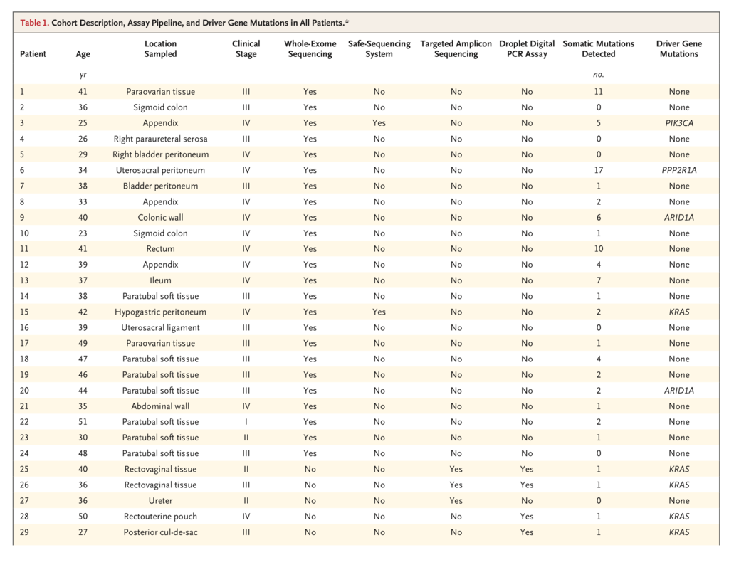
We studied nonovarian, deep infiltrating endometriosis lesions from 39 women with a mean age of 37 years. Independent molecular genetic analyses were conducted in parallel and included unbiased whole-exome sequencing in one set of lesions, with validation of driver mutations by Safe-SeqS and immunohistochemical analysis; quadruplicate targeted panel-based sequencing confirmed by a droplet digital PCR assay in a second set of lesions; and droplet digital PCR analysis alone in a third set of lesions (Table 1TABLE 1
Cohort Description, Assay Pipeline, and Driver Gene Mutations in All Patients., and Fig. S1 in Supplementary Appendix 1). The analyses carried out in the first two sets were designed and undertaken without knowledge of the results of the concurrent parallel experiments.
Exomewide Analysis of Endometriotic Lesions
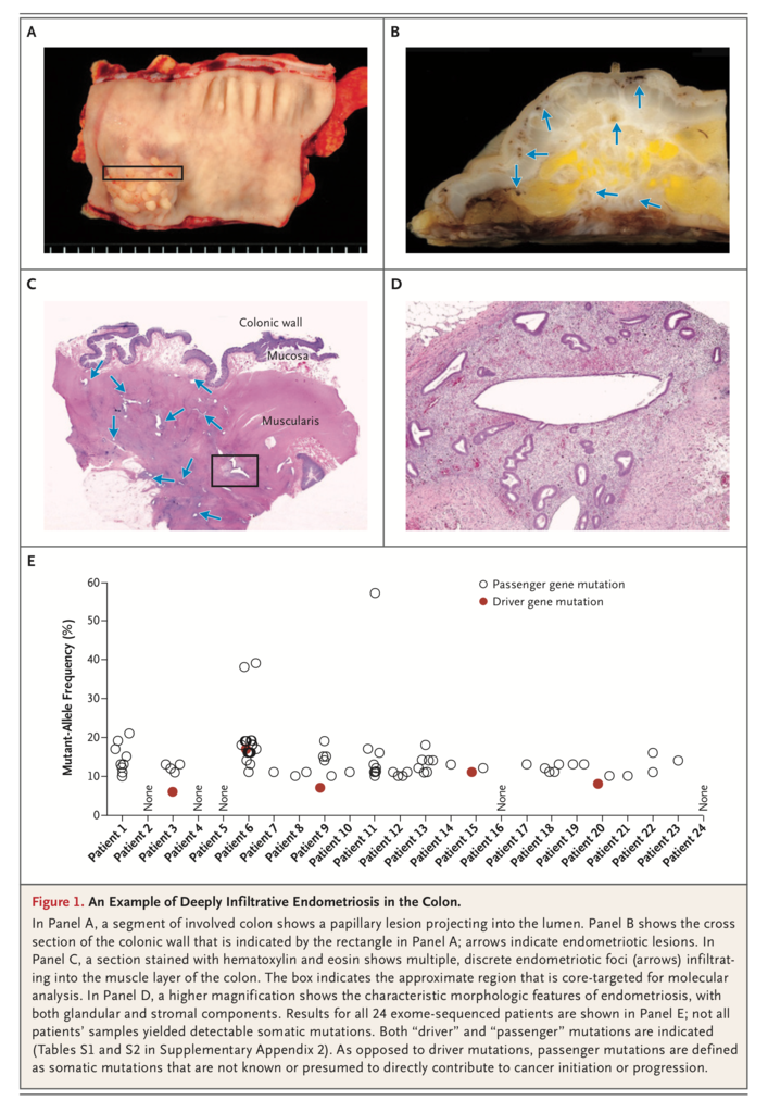
Whole-exome sequencing was performed on samples from 24 patients (Patients 1 through 24) with the use of DNA isolated from endometriosis tissue cores and matched normal tissue (Table 1 and Figure 1). Coverage was on average 104× (range, 30 to 161) in endometriosis tissue cores and 53× (range, 26 to 97) in normal tissues (Table S1 in Supplementary Appendix 2). We identified 80 nonsynonymous, somatic mutations, including 61 missense and 5 nonsense mutations, 5 insertion–deletion mutations (indels) producing frameshifts, 7 indels that were in-frame, and 2 mutations at canonical splice sites (Table S2 in Supplementary Appendix 2). The number of mutations per lesion varied widely (from 0 to 17), with a mean of 3.3 mutations per lesion (Table 1); five lesions had no detectable mutations. The mutant-allele frequencies of somatic mutations were generally low (<20%) (Figure 1E, and Table S2 in Supplementary Appendix 2), suggesting that only a subset of cells harbored mutations.
Five lesions harbored somatic mutations in cancer driver genes. Two lesions harbored frameshift inactivating mutations in the tumor suppressor ARID1A(p.L2253Cfs*14 in Patient 9 and p.G276Efs*87 in Patient 20), and hotspot activating mutations in PIK3CA (c.3127A→G; p.M1043V), KRAS (c.35G→T; p.G12V), and PPP2R1A (c.767C→T; p.S256F) were each found in one lesion. The remaining mutations were in genes that are not suspected to be driver genes in cancer.
The mutant-allele frequencies of alterations that were identified in cancer driver genes such as ARID1A, PIK3CA, KRAS, and PPP2R1A ranged from 6% to 17% and did not differ significantly from those of non–cancer driver genes. Exome data were also used to estimate DNA copy number; however, small copy-number gains were detected only in Patient 10 (Table S3 in Supplementary Appendix 2).
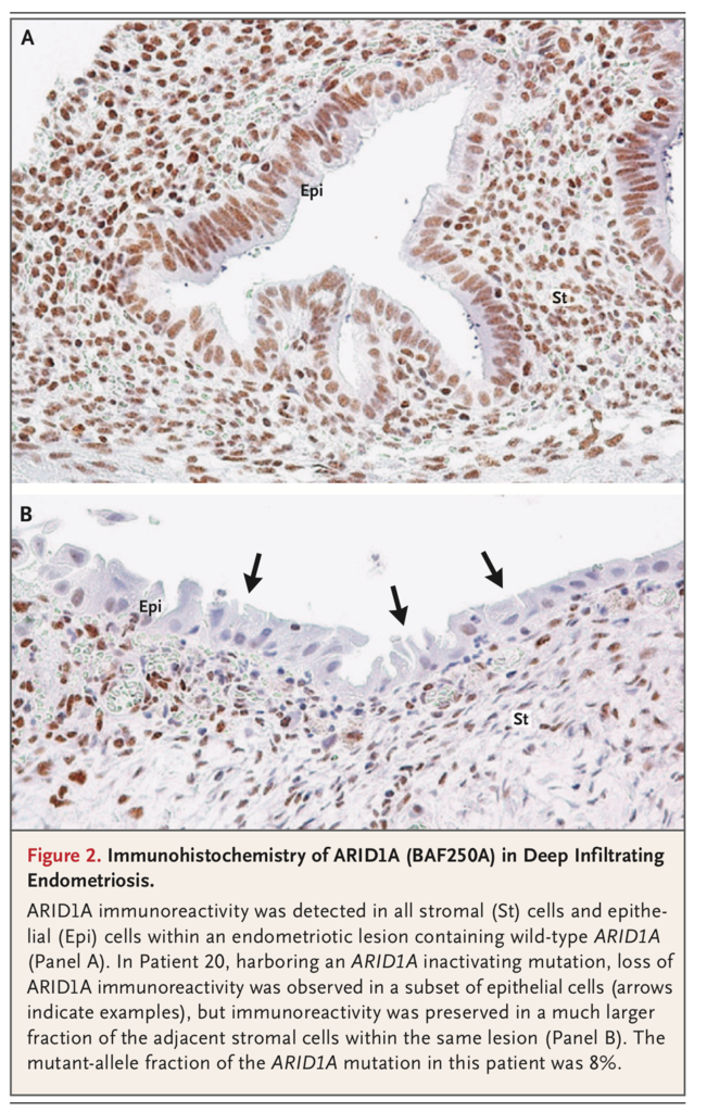
Sufficient DNA was available to validate a subset of these mutations from the same samples with the use of an orthogonal digital genomic method, Safe-SeqS.24 Safe-SeqS confirmed the existence of the somatic mutations and confirmed the allele frequencies from the original exome data (Table S4 in Supplementary Appendix 2). The hotspot KRAS mutation (in Patient 15) was further characterized by laser-capture microdissection of endometriotic lesions that separated epithelial and stromal compartments. Herein, the mutation was present in the epithelium but not in the endometrial-type stroma, a finding consistent with the low allelic fraction when both compartments were analyzed together (Table S4 in Supplementary Appendix 2). Loss of ARID1A immunoreactivity served as a surrogate for an inactivating mutation in Patient 20: ARID1A immunoreactivity was undetectable in a subset of the epithelial cells but was present in stromal cells (Figure 2). In addition, the PPP2R1A mutation in Patient 6 was validated in the epithelial compartment by a droplet digital PCR assay (Table S7 in Supplementary Appendix 2); however, we were unable to recover a pure stromal sample from the remaining lesion.
Cancer Driver Deep-Sequencing Analysis of Endometriotic Lesions
In an independent set of experiments, we analyzed tissue samples from three patients (Patients 25, 26, and 27) using two targeted panels with overlapping coverage of the hotspot regions of five genes (NRAS, BRAF, FGFFR2, HRAS, and KRAS) and the full coding sequence of six genes (PIK3CA, PTEN, TP53, CDKN2A, CTNNB1, and FBXW7) (Table S5 in Supplementary Appendix 2). Biphasic endometriotic lesions were isolated by laser-capture microdissection, and mutations were considered to be confirmed if they were present in four panel libraries for each endometriosis sample and absent from (patient-matched) germline DNA (Table S6 in Supplementary Appendix 2 and Fig. S1 in Supplementary Appendix 1). Of the three patients, two harbored activating mutations in KRAS; however, no other somatic alterations passed our filter (i.e., were present in all four libraries). Patient 25 harbored the c.35G→T (p.G12V) and c.35G→C (p.G12A) KRAS mutations, which were present at mean allele frequencies of 11.2% and 8.6%, respectively. Patient 26 had a single c.35G→A (p.G12D) KRAS mutation present at a mean allele frequency of 10.4% (Fig. S2 in Supplementary Appendix 1).
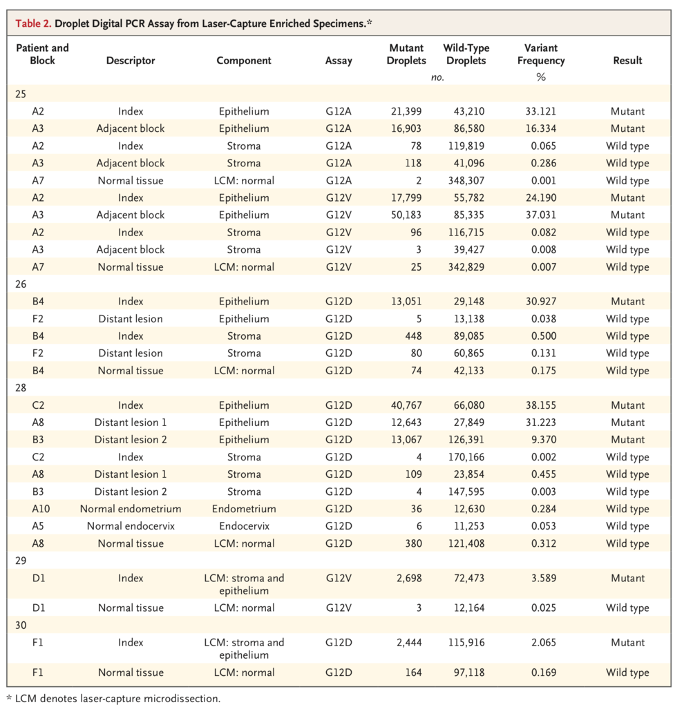
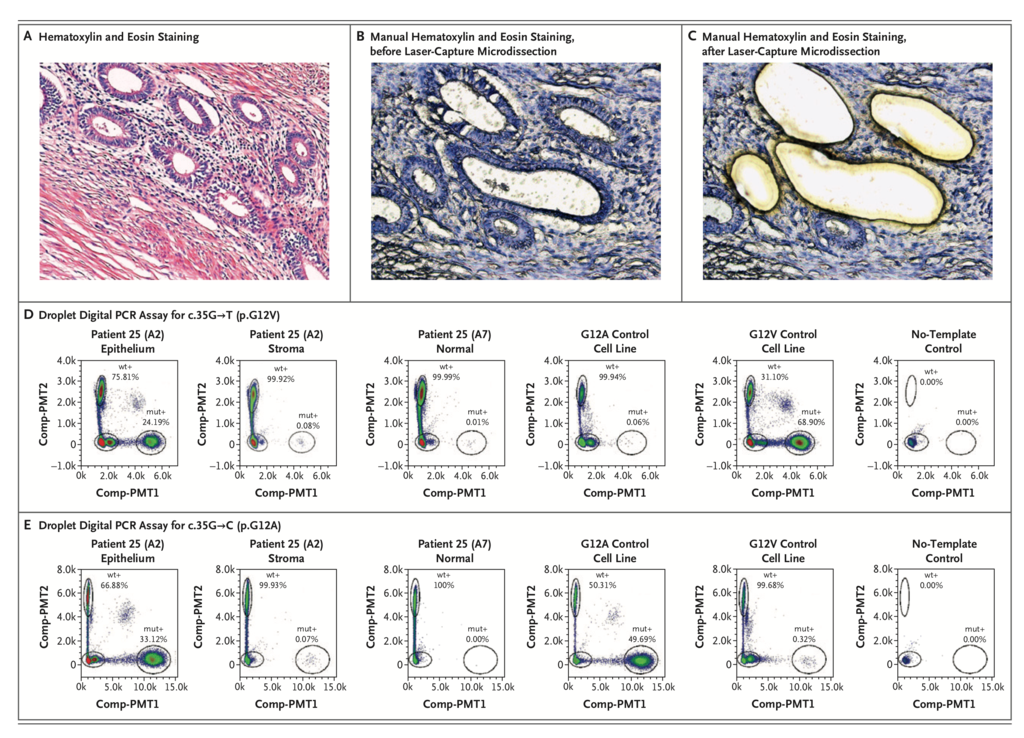
KRAS variants were validated orthogonally by droplet digital PCR assay. Two adjacent blocks from a single lesion in Patient 25 were assayed for both c.35G→T (p.G12V) and c.35G→C (p.G12A); the assay confirmed the presence of the two distinct mutations in the same endometriotic epithelial compartment: the c.35G→T (p.G12V) mutation was present in index and adjacent blocks at frequencies of 24% and 37%, respectively, and the c.35G→C (p.G12A) mutation was present at frequencies of 33% and 16%, respectively (Table 2 and Figure 3). We examined two blocks in Patient 26, each from a distinct and anatomically separated endometriotic lesion. The KRAS c.35G→A (p.G12D) mutation was confirmed in the epithelial compartment of only the index tissue block (originally subjected to panel sequencing) at a frequency of 31% (Table 2, and Fig. S2 in Supplementary Appendix 1); it was not detected in the other lesion from this patient.
Recurrent KRAS G12 Variants in Deep Infiltrating Endometriosis
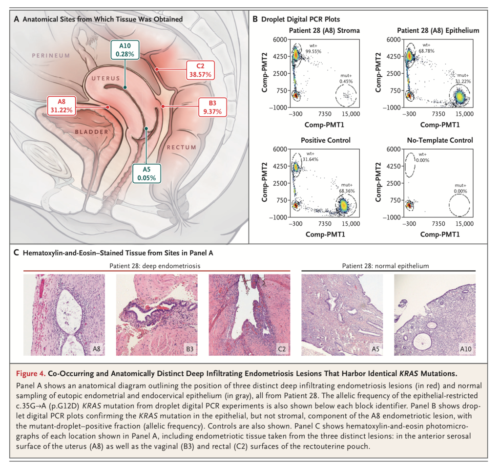
Because KRAS alterations were detected in the above two cohorts (3 of 27 patients), we analyzed deep infiltrating endometriosis lesions from an additional 12 patients (Patients 28 through 39) (Table 1) using droplet digital PCR assays for five KRAS codon 12 variants (c.35G→A [p.G12D], c.35G→T [p.G12V], c.35G→C [p.G12A], c.34G→C [p.G12R], and C.34G→T [p.G12C]). Droplet digital PCR assays were initially performed in manually macrodissected lesions (Table S7 in Supplementary Appendix 2), and lesions that showed positivity were then laser-capture microdissected, with stromal and epithelial compartments separated when possible. We detected KRAS mutations in 3 of 12 patients (25%) (Table 2). For Patient 28 with a c.35G→A (p.G12D) mutation, three independent deep lesions and normal endometrial epithelium were available for laser-capture microdissection and high-resolution droplet digital PCR analysis (Figure 4). All three anatomically distinct endometriosis lesions harbored the same KRAS c.35G→A alteration in the glandular epithelium; however, we were not able to detect the mutation in endometriosis stroma or normal eutopic endometrial epithelium from the same patient.
DISCUSSION
Despite the high prevalence of endometriosis and its effect on women’s health-related quality of life,29 very little is known about the biologic processes that underpin it, although more than a century has elapsed since this disease was first described.30,31 There is a socioeconomic imperative for endometriosis research; in the United States alone, the economic burdens are more than $12,000 per hospital stay,32 more than $11,000 per year per woman with the disorder,32,33 and more than $54 billion per year in total.34 In this study, we used a combination of next-generation sequencing and validation through highly sensitive digital genomic assays and found that the majority of benign deep infiltrating endometriosis lesions harbored somatic mutations, including mutations in the well-known cancer driver genes ARID1A, PIK3CA, KRAS, and PPP2R1A.35-37
Our reporting of these driver gene mutations in the affected patients should be considered conservative, because methods for sequencing ultra-low-input and formalin-fixed materials are still developing. Should a more complete genomic or epigenomic analysis be applied, additional driver mutations may be uncovered. Nonetheless, the driver genes that we observed to be altered are frequently mutated in endometriosis-related ovarian neoplasms and in (clonally related) adjacent and distant endometriotic lesions from patients with endometriosis-related ovarian neoplasms.14,38Yet, it is critical to note that endometriosis is generally regarded to be a nonmalignant inflammatory condition.3 The finding of cancer-associated mutations in endometriotic lesions without concurrent cancer and, in particular, in nonovarian deep infiltrating lesions that rarely (if ever) transform into cancer was surprising. These mutations may be an intrinsic characteristic of deep infiltrating endometriosis and raise interesting questions about the pathobiology of endometriosis.
In the past, studies that examined endometriosis-related ovarian neoplasms with concurrent endometriosis suggested that driver mutations shared by both lesions were the mutations responsible for the progression of the endometriosis to cancer.16,17,39,40 However, given an estimated rate of malignant transformation for endometriosis close to 1%,41 our results suggest that the presence of driver mutations alone is neither sufficient to drive the transformation of endometriosis nor indicative of likely progression to cancer. At least one study of a mouse model of endometriosis has already provided evidence that (subclonal) activating Kras mutations can sustain endometriosis but are not sufficient for malignant transformation.42 Our data also agree with studies of other organ systems that have shown cancer driver mutations in benign lesions and normal tissues.43-46 Beyond this, because only a minority of our patients harbored detectable driver mutations, the current study may suggest that driver mutations are not required for the development of (deep infiltrating) endometriosis. Perhaps more important, our findings challenge past research, in which endometriosis was typically studied in conjunction with endometriosis-related ovarian neoplasms and in which candidate single-gene approaches were used; these types of studies are unlikely to be sufficient in addressing the risk of transformation.
The probability that 5 mutations, out of the 80 mutations found, were in a pool of 125 driver genes was calculated to be P=0.001 (binomial test), which suggests that these mutations were highly unlikely to have arisen by chance alone. Because cancer driver mutations were present only in the epithelium but not the stroma of the same endometriosis lesion, we can assume that the observed mutations provide some key selective advantage to endometriotic epithelial cells. This apparent selective pressure in the epithelial compartment could result in the emergence of distinct clonal populations within the same lesion. For example, Patient 25 harbored two different KRAS codon 12 mutations. Again, not all lesions harbored detectable driver gene mutations; future studies are necessary to more accurately determine the prevalence of specific driver events, as well as to address whether cases of endometriosis without driver mutations have functionally equivalent genomic or epigenomic events that were not assessed in this study and whether mutation-harboring endometriosis represents a distinct pathology. Finally, not all lesions in a given patient were found to harbor a detectable (driver) mutation (e.g., Patient 26), a trend that has also been observed in the context of endometriotic lesions co-occurring with cancer14; these findings potentially support the coexistence of multiple endometriosis lineages within the same patient.
Deep infiltrating endometriosis can invade visceral organs and distort local anatomy, whereas in the superficial subtype of endometriosis, lesions are noninvasive and anatomical relationships are maintained. The biologic processes underpinning these distinct phenotypes is unknown. Our findings challenge the current understanding of this invasive subtype of endometriosis and open the discussion on whether deep infiltrating endometriosis can be considered as a benign neoplasm. One patient (Patient 28) had the same KRAS mutation in three spatially distinct lesions, raising the possibility that some sites of benign deep infiltrating endometriosis arise through the neoplastic process of metastasis. However, several mechanisms should be considered as explanations for this observation. First, mutations may have arisen independently of each other. However, it is unlikely that exactly the same mutation occurred independently, by chance, in all three lesions. Second, the mutation may have arisen in a single lesion and subsequently disseminated to other sites. This possibility supports the “metastasis” of somatically altered endometriosis epithelial cells but does not specifically address the contentious issue of endometriosis originating through metaplasia or dissemination from the endometrium. Nonetheless, if we choose to accept this explanation, there is also the nonmutant stromal component of the endometriosis to consider; either this component must be required to maintain fitness of the epithelial cells and therefore co-migrates (is recruited) with the epithelium during the pathogenesis of endometriosis, or it develops after the establishment of the (mutant) endometrial epithelial cell through metaplasia of nonendometrial stromal cells. Lastly, a bipotent (uterine) stem cell origin leading to both stromal and epithelial components of endometriosis has been proposed.47,48 A stem-cell–related theory may provide a plausible route for single-cell metastasis; however, this would require a specific pressure for only the epithelial progeny to gain (or retain) the cancer driver mutations that we have observed thus far. Clearly, this hypothesis requires additional validation, but it may be more compatible with rare and distant sites of endometriosis (e.g., in the lung, spine, and brain).49
A spectrum of molecular analyses are needed to address key questions about the pathogenesis of endometriosis and its clinical behavior (Table S8 in Supplementary Appendix 2). The current clinical classification of endometriosis is neither biologically based nor strongly predictive of clinical behavior50; herein lies an opportunity to improve or redesign a biologically informed classification scheme. There is a broad range of challenges faced by women with endometriosis and their caregivers; this study provides a strong rationale and the molecular foundations for the extensive characterization of all endometriosis subtypes. Given the wide range and often subjective nature (e.g., self-reported pain) of clinical characteristics across patients with endometriosis, large and well-annotated cohorts will be needed.
In summary, although endometriosis is considered to be a benign disorder from both a clinical and a histopathological perspective, well-known cancer-associated somatic mutations were found in the glandular epithelium of some deep infiltrating endometriosis lesions. These findings create new opportunities for a more detailed examination of all forms of endometriosis with the use of research approaches that are common in the study of cancer.






 留言列表
留言列表
 線上藥物查詢