Dermatology. 2017;233(1):86-93. doi: 10.1159/000468149.
Abstract
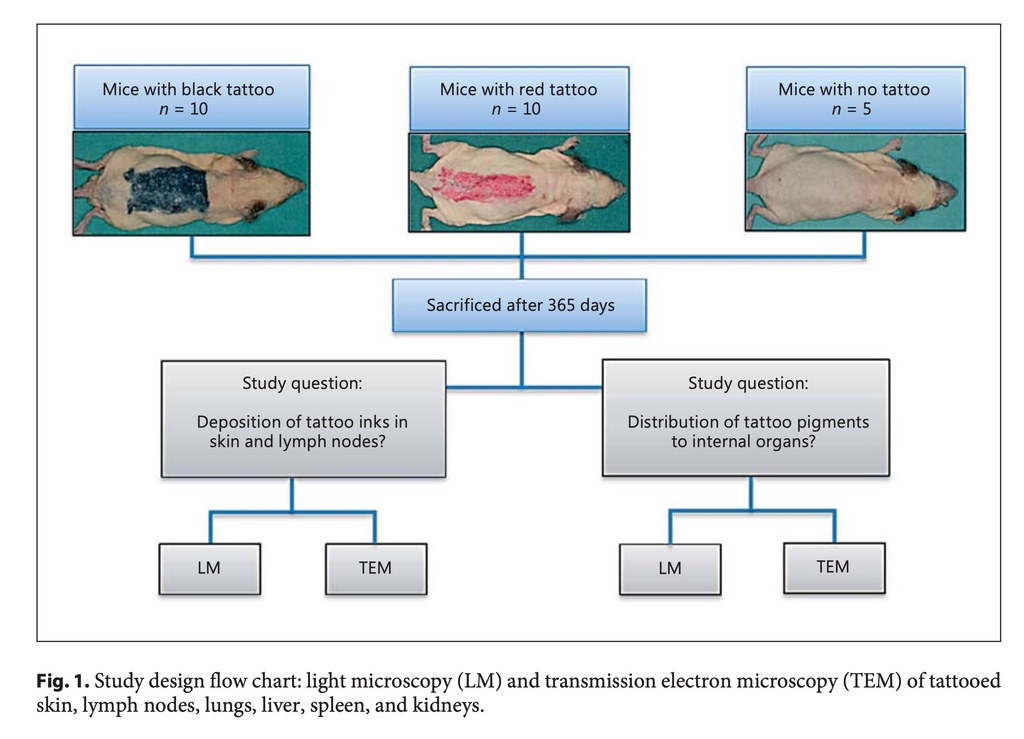
Aim: Tattoo pigments are deposited in the skin and known to distribute to regional lymph nodes. Tattoo pigments are small particles and may be hypothesized to reach the blood stream and become distributed to peripheral organs. This has not been studied in the past. The aim of the study was to trace tattoo pigments in internal organs in mice extensively tattooed with 2 different tattoo ink products. Material/Methods: Three groups of mice were studied, i.e., 10 tattooed black, 10 tattooed red, and 5 untreated controls. They were tattooed on the entire back with commercial tattoo inks, black and red. Mice were sacrificed after 1 year. Samples were isolated from tattooed skin, lymph nodes, liver, spleen, kidney, and lung. Samples were examined for deposits of tattoo pigments by light microscopy and transmission electron microscopy (TEM). Results: TEM identified intracellular tattoo pigments in the skin and in lymph nodes. TEM in both groups of tattooed mice showed tattoo pigment deposits in the Kupffer cells in the liver, which is a new observation. TEM detected no pigment in other internal organs. Light microscopy showed dense pigment in the skin and in lymph nodes but not in internal organs. Conclusion: The study demonstrated black and red tattoo pigment deposits in the liver; thus, tattoo pigment distributed from the tattooed skin via the blood stream to this important organ of detoxification. The finding adds a new dimension to tattoo pigment distribution in the body, i.e., as observed via the blood in addition to the lymphatic pathway.
Results
All mice survived the initial tattooing. Tattooing with both inks resulted in needle trauma with acute cutaneous inflammation and occasionally dermal haemorrhage. After 14 days, the skin had fully recovered in all tattooed mice. The mice did not present any noticeable general or internal disease during the study, and there were no unusual drop-outs from the study.
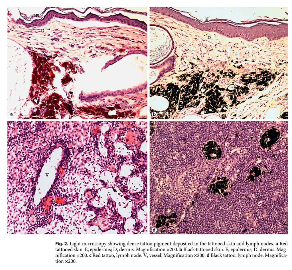
The presence of ink in the skin and lymph nodes was confirmed visually and by histology after the mice had been sacrificed. Histology confirmed the deposition of ink primarily in the dermis.
Transmission Electron Microscopy
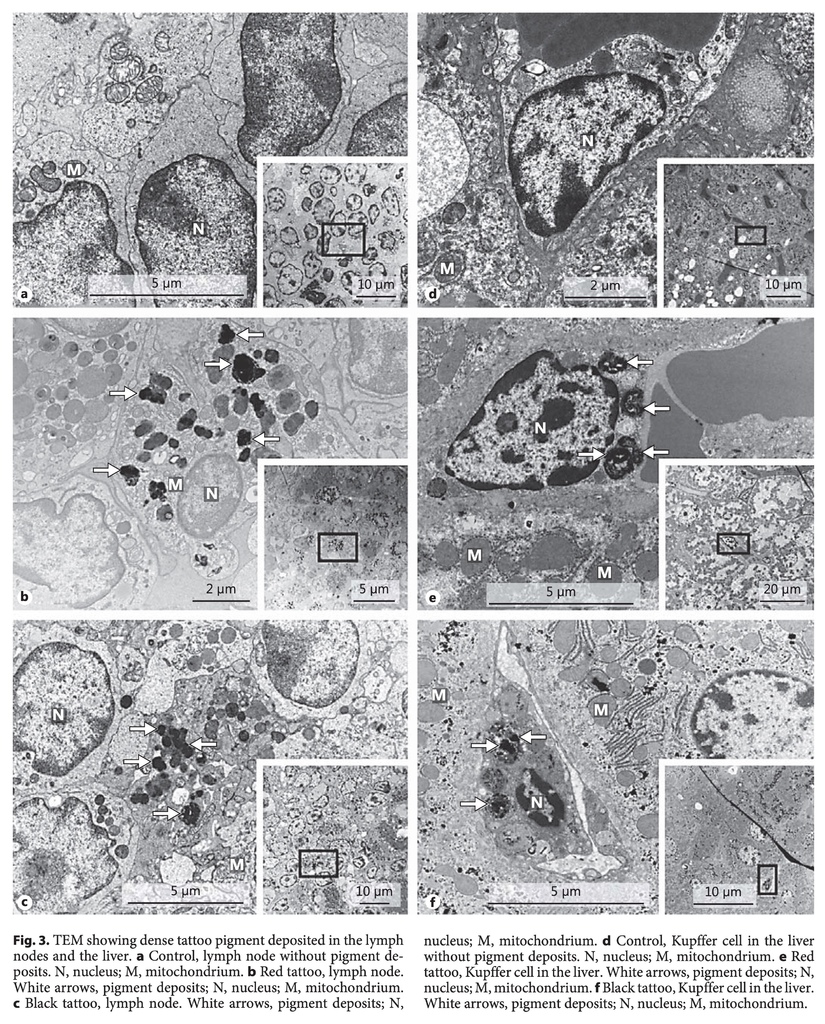
All 6 organs (skin, lymph nodes, lungs, liver, spleen, and kidneys) from the 20 tattooed mice and the 5 controls were investigated with TEM. In the skin, lymph nodes, and the liver we observed electron-dense deposits indicating potential tattoo pigment. In the lymph nodes we observed a diffuse pattern of highly electron-dense material distributed in cells with an ultrastructure resembling macrophages (Fig. 3b, c). This observation was made in 18 of the 20 tattooed mice, and no similar intracellular electron-dense deposits were observed in the controls (Fig. 3a). In the liver, we observed the same kind of electron-dense deposits in membrane-bound organelles, specifically located in the Kupffer cells (Fig. 3e, f). In 19 of 20 samples from the liver of the tattooed mice such electron-dense material was observed. No similar electron-dense deposits were found in the untreated controls without tattoos (Fig. 3d). Concerning other internal organs, e.g., the kidney, spleen, and lung, no electron-dense deposits were detected in any of the 3 groups of mice studied. Accordingly, pigment deposition in internal organs was exclusively observed in the Kupffer cells of the liver in addition to their presence in the skin and lymph nodes.
Discussion
The study made the original and direct observation that tattoo pigment originating from the tattooed skin is observed in the liver. Tattoo pigment deposits in a distant organ such as the liver can only be explained by a blood-borne distribution.
Previous studies of the systemic distribution of nanoparticles in mice and rats were performed using gold and silver particles and quantum dots, which are detectable in internal organs in small amounts by microscopic methods and special high-sensitivity techniques such as inductivity-coupled plasma mass spectrometry [16,17,18]. Following intradermal injection into the dorsal flank of mice, Gopee et al. [16] observed quantum dot deposits in the liver, regional draining lymph nodes, kidney, spleen, and hepatic lymph nodes. Following subcutaneous injection in rats, Tang et al. [17] found silver nanoparticles in the kidney, liver, spleen, brain, and lung. Silver nanoparticles induced blood-brain barrier destruction, astrocyte swelling, and caused neuronal degeneration. Following intratracheal insertion in mice, Sadauskas et al. [18] found gold particles in the liver. They injected nanoparticles in the size range of 2-100 nm and found the translocation rate greatest with the 2-nm gold particles. Kreyling et al. [19,20] found that translocation of nanoparticles studied in rats depended on particle size and additionally chemical composition, shape, electrical charge, and other features. Accordingly, there is experimental evidence from various small animal studies that injection into the skin and other local exposures to nanoparticles are subject to blood-borne distribution to major organs and even to distant sites with a known special physical and chemical translocation barrier, such as the brain.
We aimed to perform an authentic study addressing the systemic distribution of tattoo pigments. We used two commercial tattoo ink stock products commonly used by tattooists, a black and a red ink. Carbon black and red azo pigments are sized nanometres or micrometres and difficult to detect in tissues in small amounts as compared to experimental particles of predefined size and characteristics facilitating their detection.
By light microscopy we observed large aggregates of pigment in the skin and lymph nodes in accordance with the literature [6,7,8,9,21]. Light microscopy includes a much larger tissue sample and section thickness but this method is limited by inadequate resolution and only allows larger aggregates of pigments to be visualized.
Accordingly, this method could not detect pigment in internal organs because of the limited resolution, or since pigment was truly absent.
By TEM we found particle deposits in the liver with both black and red pigments, in contrast to no detectable pigment in other organs studied, e.g., the lung, spleen, and kidney. Samples and sections for TEM only represent a minute element of the complete organ, and deposits can occur sporadically in the organ without being included in the particular sample. Accordingly, TEM may have underestimated or overlooked deposits of pigments in organs with minimal deposition. The two pigments examined have different size and chemical characteristics but, nevertheless, distributed to the same degree to the liver.
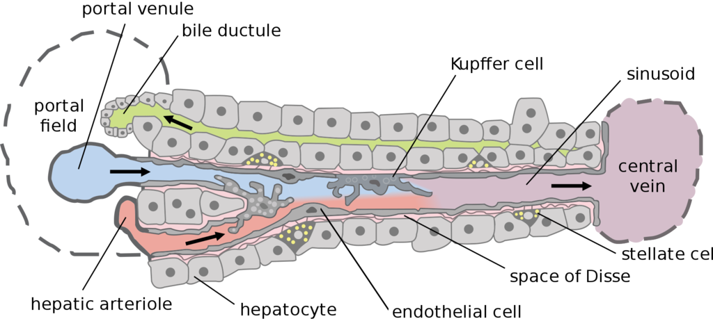
Deposition in the liver was observed in the Kupffer cells. Kupffer cells, also known as stellate macrophages and Kupffer-Browicz cells, are resident and specialized macrophages of the liver, lining the walls of the sinusoids, and have a gatekeeper function. They are critical components in the phagocytic system and play a central role in the hepatic and systemic response to pathogens [22,23,24]. Kupffer cells are part of the detoxification system of the body and serve to encapsulate and inactivate particulate elements, which have reached the blood and passed through the liver. Accordingly, it is reasonable that we detected tattoo pigment particles especially in this cell parallel to the finding of tattoo pigments in macrophages of the skin and in lymph nodes.
Previous studies demonstrated that tattooed skin and regional lymph nodes hold large amounts of tattoo pigment [4,25]. A study of human skin tattooed black and the regional lymph nodes confirmed that black pigment and polyaromatic hydrocarbons were found in the lymph nodes in high amounts [21]. Deposition of tattoo pigment in the skin is not stationary. In a study of mice tattooed with Pigment Red 22, Engel et al. [26] showed that after 42 days the amount of pigment in the skin had decreased by 32%. This was done by quantitatively extracting the pigment at different times after tattooing. Furthermore, clearance of the skin from tattoo pigment was shown to depend on light exposure and photodecomposition [26]. Thus, distribution of tattoo pigment from skin to the regional lymph nodes is significant and occurs dynamically, especially during the first few weeks after tattooing when systemic distribution might occur [27].
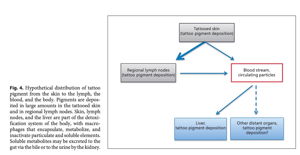
Tattoo pigment may reach the blood either by translocation directly from the skin to the circulation or indirectly by release from the lymph nodes as part of normal lymphatic drainage. We showed for the first time that tattoo pigments could be observed in the liver. This can only be explained by their temporary circulation in the blood. It remains unknown whether circulating pigment originates from capillary blood of the skin or from the lymph or from both sources. Our hypothesis on tattoo pigment distribution from the skin to the body is shown in Figure 4.
Taking small animal experiments and the study of experimental nanoparticles into consideration, optimizing detection, it is likely that we may have overlooked tattoo pigment deposits in other internal organs [16,17,18]. This will require a dedicated experimental set-up using another technique, as exemplified by the study of organ distribution of radiolabelled tattoo pigments.
Clinical records of tattooed humans have not reported any conditions of the liver leading to any identified disease of the liver caused by tattoo pigments [28,29]. Thus, our finding appears not to have any known clinical correlate. Finding the tattoo pigment in macrophages and Kupffer cells indicates that the body encapsulates and detoxifies tattoo pigment using known physiological defence mechanisms. Accordingly, the risk and concern related to tattoo pigment deposits in the periphery might be overestimated. It has to be considered that tattoos are essentially permanent markers of skin already after 1 single-dose exposure. Risk estimation by the equation (risk = hazard × exposure) would speak in favour of a limited long-term risk of tattooing since exposure is not repeated or continuous.
The toxicology of tattoo inks and particles from industrial production remains highly complex and a constant matter of concern [28]. It is unknown whether specific distant organs may have a special affinity for tattoo pigment deposition and thus may be subject to rare events and hazards not yet identified. Distant organs may also be especially vulnerable to pigment particles. For comparison, it was a very late notice when it became apparent that metal-on-metal total hip arthroplasty continuously released metallic particles to the blood that were shown to result in particle deposits in the brain causing neurological symptoms [30,31]
Our new finding of tattoo pigments released from the tattooed skin, circulating in the blood and observed directly in the liver introduces an unexplored risk scenario of potential systemic adverse events of different kinds affecting specific organs.




 留言列表
留言列表
 線上藥物查詢