常常聽到人家說要看看綠色,尤其是遠方的綠色,對眼睛比較好,但是,為何不是看其他顏色呢?
這件事情要講,就要先講一種病,稱為黃斑部病變:
簡單的說:視網膜就像是照相機的底片存在於眼睛裡,其中黃斑部乃是視功能最敏感的地區,因含黃色素,稱為黃斑部。
看圖:
在看下去之前,先學一個名詞: retinal pigment epithelium (RPE)

The area within the outer white circle (indicating a retinal diameter of about 6 mm) is the macula lutea, which is Latin for yellow spot or stain. The inner circle (diameter, 0.8 mm) borders the fovea, which is the central pit of the macula, where the preponderance of cones over rods is highest for sharp vision. The graph superimposed on this normal fundus shows the drop in attainable visual acuity in relation to the distance from the fovea. The effect on visual acuity of the location and area covered by a central age-related macular degeneration scar can be estimated on the basis of this measure. The retina, which has 10 layers, is the inner lining at the back of the eyeball. The inset shows the two outer layers that contain the purple light-sensitive rod and cone photoreceptor cells supported by Müller cells, all embedded in the interphotoreceptor matrix, in close contact with the retinal pigment epithelium (RPE). The RPE is surrounded by two extracellular matrixes, the interphotoreceptor matrix and Bruch's membrane. Between the RPE and the outer wall of the eye (sclera) are Bruch's membrane, the choriocapillaris, and a larger vessel layer, the choroid. Ruysch's complex includes the RPE, Bruch's membrane, and the choriocapillaris.
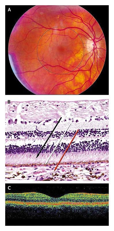
Panel A is a photograph of a normal fundus and retina. The area within the black circle is the macula. Panel B shows a histologic section of normal retina, with photoreceptors (black arrow), retinal pigment epithelium (white arrow), and the choroid (red arrow). (Image courtesy of Mort Smith, M.D.) Panel C shows a cross-sectional image of the retina generated by optical coherence tomography, an imaging technique that allows for real-time, noninvasive visualization of retinal architecture.
但是當黃斑部受到藍光傷害時,就會漸漸的受損,當然,隨著年齡增加,你所接受到的藍光傷害也會增加,因此你也會看到這樣一個名詞:Age-Related Macular Degeneration
看一下分類:
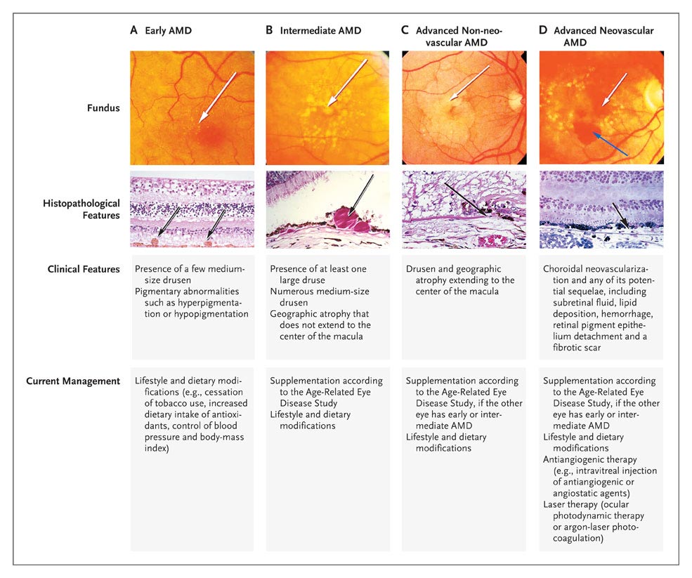
Classification of Age-Related Macular Degeneration (AMD).
Column A shows medium-size drusen (arrows) in early AMD, and Column B shows a large druse (arrows) in intermediate AMD. In Column C, a photograph of the fundus shows geographic atrophy (white arrow), and a histopathological photograph shows geographic atrophy with loss of Bruch's membrane (black arrow). In Column D, the photograph of the fundus with neovascular age-related macular degeneration shows subretinal hemorrhage (blue arrow) and choroidal neovascularization (white arrow), and the histopathological photograph shows choroidal neovascularization (black arrow). (Images courtesy of Mort Smith, M.D., and Deepak Edward, M.D.)
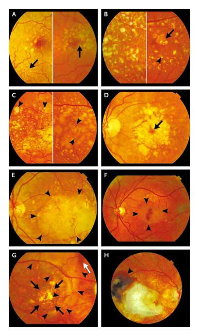
Progression from Early to Late Age-Related Macular Degeneration.
Drusen can be classified according to size, appearance, biochemical composition, and examination technique.5,6 With increasing age, drusen become confluent and larger, sometimes crystalline, less circumscribed, or accompanied by hyperpigmentations or hypopigmentations of the RPE. Panel A shows early age-related macular degeneration in two maculas: the macula shown on the left-hand side contains small drusen (arrow) and some large, indistinctly bordered drusen in the fovea; the macula shown on the right-hand side contains more drusen and focal hyperpigmentation (arrow). The left-hand side of Panel B shows early age-related macular degeneration characterized by extensive small and large drusen in and around the macula; the right-hand side shows crystalline drusen (arrowhead) and a small patch of late dry age-related macular degeneration (arrow). The left-hand side of Panel C shows early age-related macular degeneration, with crystalline and calcified drusen (arrowheads); on the right-hand side (also early age-related macular degeneration) are large confluent drusen leading to a drusenoid detachment of the RPE, with hyperpigmentation (arrowheads). Panel D (late age-related macular degeneration) shows dry age-related macular degeneration, with a central island in which photoreceptors are still functioning (arrow); this eye has a complete ring scotoma around a small central visual-field remnant. Panel E (late age-related macular degeneration) shows wet age-related macular degeneration in the form of a large serous detachment of the RPE (with borders marked by arrowheads) caused by fluid leaking from a subretinal neovascular membrane. Panel F (late age-related macular degeneration) shows the development of wet age-related macular degeneration with a subfoveal hemorrhage surrounded by detachment of the RPE (arrowheads). Panel G (late age-related macular degeneration) shows dry age-related macular degeneration (black arrows), in which the orange lines are large choroidal vessels, surrounded by glial scar tissue (arrowheads) resulting from a large subretinal hemorrhage with a small remnant (white arrow). Panel H (late age-related macular degeneration) shows cicatricial wet age-related macular degeneration, with glial scarring in the macula and remnants of hemorrhages at its temporal border (arrowhead).
看一下年齡的差別:
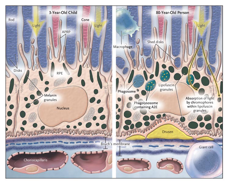
RPE Cell in a 3-Year-Old Child (Left-Hand Panel) and an 80-Year-Old Person (Right-Hand Panel).
The outer segments of the rods and cones are embedded in the interphotoreceptor matrix (blue-gray areas) and partially surrounded by apical pseudopodial RPE processes (APRP). The shed disks (right-hand panel) become encapsulated in the phagosomes and are digested in phagolysosomes in the cell cytoplasm of the RPE. Macrophages and fused macrophages (giant cells) remove cellular debris around the cell. Light-induced toxicity occurs as light is absorbed by the various chromophores in the lipofuscin granules. This damages DNA and cell membranes and causes inflammation and apoptosis. The right-hand panel shows enlarged lipofuscin granules, the thickened Bruch's membrane, and the attenuation of the choriocapillaris. The central elastic lamina in Bruch's membrane becomes more porous in old age.
好了,黃斑部病變就不再繼續解釋了,圖片來源:NEJM
Volume 358:2606-2617 Volume 355:1474-1485,有興趣自己去看更詳細的說明或是你要看這邊
所以現在要告訴大家的,就是藍光傷害,是的,根據色彩學來說,藍色和黃色是互補的
所以眼睛裡面的黃斑部,就是幫助我們吸收和承受光線中的藍光傷害,也就是偏藍色的那些光譜,像是藍色、紫色、紅色。
接著,我們要講另外一個東西:就是葉子。
沒錯,葉子,大家知道為何葉子是綠色的嗎?
依據反射光學的解釋,你看到的綠色,是反射過後的顏色,但是對於透視光學來說,你看到的綠色,就是物體吸收的顏色,當然,葉子不是透明的,所以他是將綠色反射回來給我們看到,然後吸收掉其他的顏色。
為何他會吸收其他的顏色呢?演化結果......無法解釋(葉綠素只有吸收這些長度的光線,才有最佳的轉換效率)。
但是我們知道葉子裡面存在一種東西叫做葉綠素,葉綠素本身對於兩種長度的光譜吸收最強
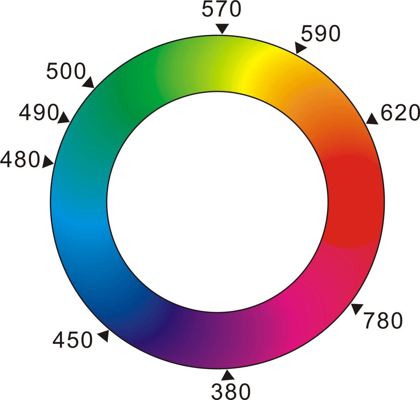
一個就是:640-660nm,另外一個就是430-450nm。看一下上面的光譜,你就可以知道,
640-660是紅色光區,430-450是藍紫色光區
也就是說,葉綠素將光線中的藍紫紅都吸收掉了,把剩下的顏色反射給我們。
也因此,當我們遠望綠色時,可以收到兩個效果:
1.調解水晶體的肌肉和水晶體本身可以得到遠距離的調節,以彌補長期近距離的疲勞
2.看綠色時可以紓緩黃斑部收到的傷害,讓黃斑部有休息的時間。
另外我找到了一篇有趣的科普概念,給大家當做參考:
光的三原色與視神經的對比傳遞




 留言列表
留言列表
 線上藥物查詢