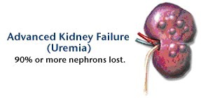
Medical progress has altered the course and thus the definition of uremia, which once encompassed all the signs and symptoms of advanced kidney failure. Hypertension due to volume overload, hypocalcemic tetany, and anemia due to erythropoietin deficiency were once considered signs of uremia but were removed from this category as their causes were discovered. Today the term “uremia” is used loosely to describe the illness accompanying kidney failure that cannot be explained by derangements in extracellular volume, inorganic ion concentrations, or lack of known renal synthetic products. We now assume that uremic illness is due largely to the accumulation of organic waste products, not all identified as yet, that are normally cleared by the kidneys.
No specific time point demarcates the onset of uremia in patients with progressive loss of kidney function. The features of uremia identified in patients with end-stage kidney failure may be present to a lesser degree in people with a glomerular filtration rate that is barely below 50% of the normal rate, which at 30 years of age ranges between 100 and 120 ml per minute per 1.73 m2 of body-surface area. Thus, in the United States alone, uremic symptoms may be present to some degree in an estimated 8 million people who have a glomerular filtration rate below 60 ml per minute per 1.73 m2 of body-surface area.1 However, early symptoms of uremia, such as fatigue, are nonspecific, making the condition difficult to identify. At present, moreover, we can slow progression to kidney failure but can treat uremia only by replacing kidney function. Thus, the question of whether a patient has uremia comes down to whether dialysis or a transplant would be beneficial.
Treatment of uremia is now dominated by dialysis, in large part because donor kidneys are in short supply. In the United States in 2004, approximately 100,000 people began receiving kidney-replacement therapy for end-stage renal disease, and 335,000 people were receiving ongoing treatment with dialysis.2 In some cases, patients are treated with dialysis for decades, but overall outcomes are disappointing. The 5-year survival rates between 1995 and 1999 were under 35% for both hemodialysis and peritoneal dialysis. Patients treated with dialysis are hospitalized on average twice a year, and their quality of life is often low.
Not all of the illness of a patient undergoing dialysis can be ascribed to uremia. Indeed, the evolution of dialysis has made the effects of uremia more difficult to distinguish, since the severity of classic uremic symptoms is attenuated. Instead, patients undergoing dialysis now have a new illness, which Depner3 aptly named the “residual syndrome.” This illness comprises partially treated uremia; ill effects of dialysis, such as fluctuation in the extracellular fluid volume and exposure to bioincompatible materials; and residual inorganic ion disturbances, including acidemia and hyperphosphatemia. In many patients, the residual syndrome is complicated by the effects of advancing age and systemic diseases that were responsible for the loss of kidney function.
Although patients undergoing dialysis have a complex illness, there are compelling reasons to believe that inadequate removal of organic wastes is an important contributor. Dialysis is initiated when uremic symptoms, among which anorexia and lethargy are usually the most prominent, advance to the point at which treatment is expected to effect an improvement. The glomerular filtration rate at this point averages about 7% of the normal value. As compared with such a low glomerular filtration rate, conventional dialysis provides only slightly better removal of many solutes and inferior removal of some. Renal replacement therapy does keep patients alive, but because of these limitations, it does not completely relieve uremic symptoms. The fact that transplantation reverses this residual syndrome constitutes strong evidence for the ill effects of toxic solute accumulation despite dialysis. Successful transplantation, which can restore the glomerular filtration rate to more than half the normal value, markedly improves the overall quality of life and enhances specific functions, including sleep, sexual function, cognition, exercise capacity, and, in children, growth.4-7
SOLUTES CLEARED BY THE KIDNEY AND RETAINED IN UREMIA
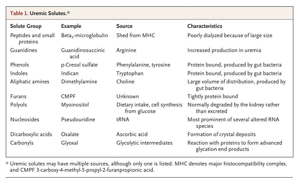
Although retained uremic solutes cause symptoms, identifying the responsible solutes has proved difficult because of the multiplicity of retained solutes (reviewed by the European Uremic Toxin Work Group8) as well as the variety and subtlety of the symptoms. Advances in chromatography and spectroscopy continue to lengthen the list of implicated solutes. In general, solutes that accumulate in the highest concentrations, and were therefore identified first, have been the most studied. But only a few compounds have been linked to specific toxic effects.9-11 Plasma concentrations of several compounds correlate more closely with altered mental function than do concentrations of urea. Some of these compounds, including certain guanidines, accumulate in the cerebrospinal fluid, which is consistent with their proposed effects on the brain.12 However, we lack experiments showing that uremic signs and symptoms can be replicated by raising solute levels in people or animals with normal kidney function to equal those in patients with uremia. Uremic solutes are therefore usually categorized on the basis of their structure. Selected examples are shown in Table 1
.
Urea is quantitatively the most important solute excreted by the kidney and was the first organic solute detected in the blood of patients with kidney failure. Both hemodialysis and peritoneal dialysis are currently prescribed to achieve target values for urea clearance. Yet early studies indicated that urea itself causes only a minor part of uremic illness.13,14 One study showed that uremic symptoms were relieved by initiation of dialysis, even when urea was added to the dialysate to maintain the blood urea nitrogen level at approximately 90 mg per deciliter (urea, 32 mmol per liter).14
Other simple nitrogen-containing solutes that accumulate in uremia include the aliphatic amines monomethylamine, dimethylamine, and trimethylamine. These compounds are produced by both gut bacteria and mammalian cells. They are positively charged at physiologic pH, and their removal during intermittent hemodialysis may be limited by their preferential distribution within the relatively acidic intracellular compartment.15 The uremic fetor, or fishy breath, of patients with uremia is attributable to trimethylamine, and amines have been associated with impaired brain function in both patients and animal models.16-18
Many uremic solutes contain aromatic rings. The uremic phenols derive from the amino acids tyrosine and phenylalanine and from aromatic compounds in vegetables. Indoles arise in analogous fashion from tryptophan and vegetable indoles. In both cases, metabolism of parent compounds by methylation, dehydroxylation, oxidation, reduction, or conjugation produces a bewildering array of solutes. In contrast to methylamines, conjugated phenols and indoles are often negatively charged and contribute to the increase in the anion gap observed in kidney failure. The structural similarity of waste phenols and indoles to neurotransmitters has encouraged speculation that these compounds interfere with the function of the central nervous system, but evidence of the toxicity of individual compounds is weak. The most extensively studied phenol is p-cresol, which is formed by colonic bacteria from tyrosine and phenylalanine and then circulates conjugated with sulfate.19,20High p-cresol levels have been associated with poor outcomes in patients undergoing dialysis.19,21
The kidney clears not only small molecules but also proteins with molecular weights between 10 and 30 kD. Plasma levels of these low-molecular-weight proteins rise as the kidney fails, but only beta2-microglobulin, which causes dialysis-related amyloidosis, has known toxicity.9,22 The extent to which kidney failure increases the levels of peptides with molecular weights between 500 D and 10 kD is less well defined,23 although it is expected that proteomic techniques may ultimately yield a more complete picture.23,24
SOLUTE REMOVAL BY DIALYSIS
Uremia could theoretically be treated by reducing solute production, but this is not part of current practice. High protein intake increases the production of many solutes, including various guanidines, indoles, and phenols. Patients with kidney failure tend to reduce their protein intake spontaneously, and before dialysis became available, physicians found that marked protein restriction relieved uremic symptoms.25,26 Protein restriction can have ill effects, however, and it is now recommended that patients undergoing dialysis receive 1.2 g of protein per kilogram of body weight per day, which is nearly the amount provided by an average diet in the United States. Since a number of the best-known uremic solutes — such as aliphatic amines, D-amino acids, methylguanidine, hippurate, and many indoles and phenols — are produced entirely or in part by gut bacteria, the use of sorbents to reduce the load of such solutes has been considered but has not been systematically studied.27
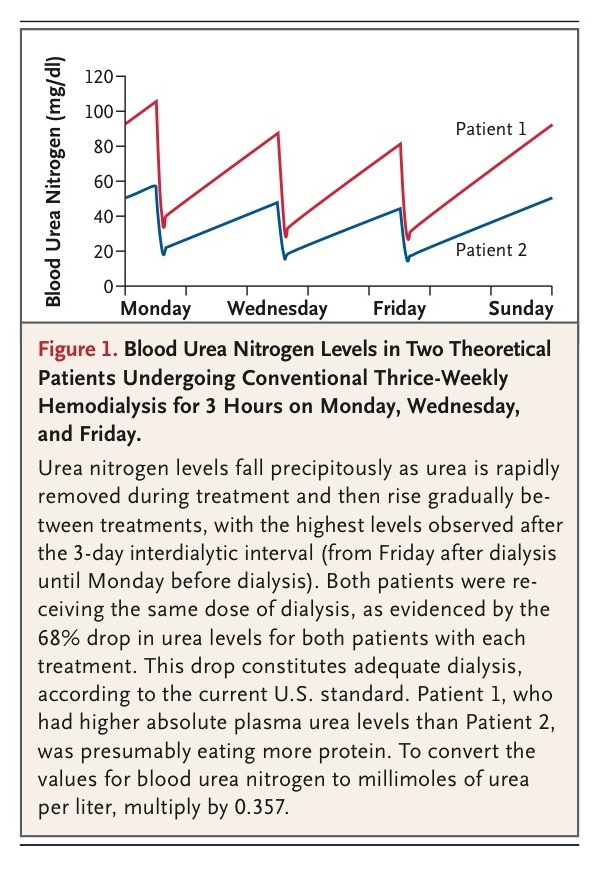
At present, most patients with end-stage renal disease undergo hemodialysis three times per week. The dialysis prescription is adjusted to remove about two thirds of the total-body urea content during each treatment (Figure 1
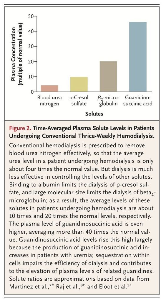
). This standard was adopted after clinical trials showed that to a point, patient outcomes improve with increasing fractional urea removal.3,28 The Hemodialysis (HEMO) Study29 showed that outcomes are not improved by increasing fractional urea removal above the current standard. Treatment that removes the majority of urea should also remove more toxic solutes if, like urea, they diffuse relatively freely throughout body water. But removal of many solutes is more limited, owing to large molecular size, protein binding, or sequestration within body compartments. Plasma levels of such solutes therefore remain much higher than normal urea levels in patients undergoing conventional dialysis (Figure 2
). Removal of such solutes can be increased by various modifications of the standard dialysis treatment. If a specific modification of dialysis reduced illness, this might reveal the characteristics of important uremic toxins.
Large Solutes
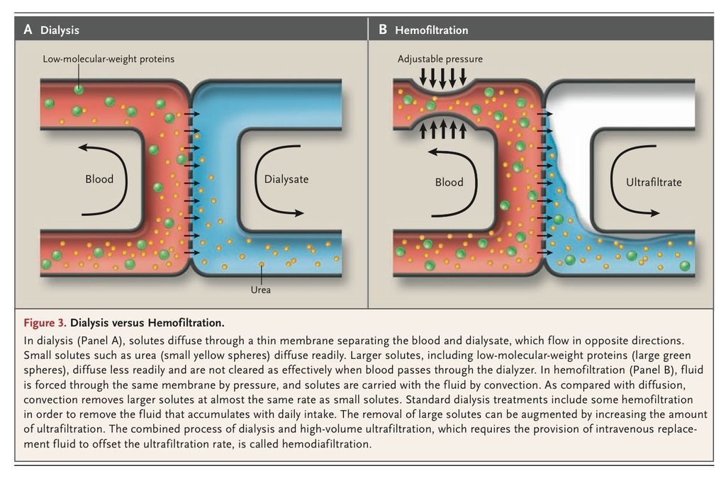
Hemodialysis was initially performed with the use of dialysis membranes that provided limited clearance of solutes with molecular weights above 1 kD. Treatment with the use of these membranes wakened comatose patients, relieved vomiting, and partially reversed other classic uremic symptoms. This provided evidence, which remains convincing, that some important uremic toxins are small. But clinical observations led pioneering investigators to speculate that other important toxins were “middle molecules,” with molecular weights between 300 D and 2 kD.32 The original middle-molecule hypothesis was never carefully tested. Although the phrase “middle molecules” remains in use, its meaning has gradually shifted to include larger solutes. The adoption of new, more permeable membrane materials essentially ended investigation of the relative toxicity of solutes in size ranges below 1 kD. However, the toxicity of solutes with molecular weights above 1 kD remains under investigation. Increasing the removal of large solutes, such as beta2-microglobulin, with the use of “high-flux” as compared with “low-flux” membranes had no significant benefit in the HEMO Study.29 However, the clearance of large molecules can be increased further by adding ultrafiltration to the dialysis process (Figure 3
). Ongoing multicenter trials may reveal whether the combination of ultrafiltration with dialysis, called hemodiafiltration, improves outcomes.33
Solutes Bound to Albumin
Solutes that bind to albumin are poorly removed by conventional dialysis3,20,34,35 not because they are large but because only the free, unbound solute concentration contributes to the gradient driving solute across the dialysis membrane. When expressed as multiples of normal levels, the levels of these compounds are therefore higher than those of unbound solutes, such as urea, in patients undergoing hemodialysis. There is reason to suspect that at least some protein-bound solutes are toxic. The normal kidney clears many protein-bound solutes by active tubular secretion. Presumably, the combination of protein binding and tubular secretion represents an evolutionary adaptation that allows the excretion of toxic molecules and keeps the extracellular fluid concentrations of their unbound fraction very low. The aggregate toxicity of protein-bound solutes could theoretically be assessed by comparing the effects of different modifications of dialysis, but this has not been attempted in practice.36
Sequestered Solutes
Other solutes are sequestered, or retained, in compartments where their concentration does not equilibrate rapidly with that of the plasma.37 Intermittent dialysis may rapidly lower the plasma concentration of such solutes but removes only a small portion of the total-body solute content. Sequestration only slightly impairs the removal of urea, which is currently used to assess the adequacy of dialysis.3 But other solutes may equilibrate more slowly than urea between body compartments, such as between cell water and plasma, effectively sequestering them from dialysis. Only a few solutes have been carefully studied, but clinically significant sequestration has been demonstrated for phosphate, creatinine, uric acid, several guanidines, and beta2-microglobulin. Theoretically, the contribution of sequestered solutes to uremic toxicity, like the contribution of large solutes or protein-bound solutes, could be assessed by comparing the efficacy of different dialysis prescriptions. When treatment is intermittent, the removal of sequestered as compared with rapidly equilibrating solutes can be increased by lengthening each dialysis session or by increasing the number of sessions per week. Retrospective studies have shown that longer sessions are associated with better outcomes, but these studies were not sufficiently well controlled to confirm that lengthening treatment is beneficial.38 In the United States, the combined preference of patients and providers for short dialysis sessions has reduced treatment time toward the minimum required to achieve the target urea removal, making the average duration of each hemodialysis session about 3.5 hours.
SIGNS AND SYMPTOMS OF UREMIA
Frequently identified signs and symptoms of uremia are listed in Table 2
. That a fundamental metabolic disturbance such as uremia should have such a wide variety of consequences is not remarkable. The complications of untreated diabetes and hyperthyroidism are similarly extensive. But uremia is different in that we cannot trace all its complications to dysregulation of a single, key compound. And with the exception of kidney transplantation, current therapy for uremia is less successful than insulin and thyroid hormone replacement in restoring normal function.
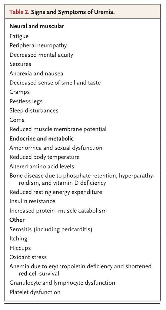
Given the signs and symptoms listed in Table 2, it is not surprising that the quality of life declines in people with chronic kidney disease. A panel preparing treatment guidelines concluded that well-being was reduced when the glomerular filtration rate was less than 60 ml per minute per 1.73 m2 of body-surface area.39 Subjects in the Modification of Diet in Renal Disease study who had glomerular filtration rates of less than 55 ml per minute per 1.73 m2 reported fatigue and reduced stamina that correlated with this rate.40 In a study using the Medical Outcomes Study 36-item Short-Form General Health Survey, people who had a glomerular filtration rate of less than 50 ml per minute per 1.73 m2 but who were not yet being treated with dialysis scored lower than the general population on 8 of the instrument's 10 scales.41 Physical functioning is also often impaired in patients with kidney failure. In those undergoing dialysis, exercise capacity has been reported to average only about 50% of normal capacity. Treatment of anemia improves but does not normalize exercise capacity. The most detailed studies suggest that susceptibility to fatigue is attributable to both muscle energy failure and neural defects.42 The degree to which the quality of life is impaired by the uremic environment, by the deconditioning of the patient, and by the effects of coexisting conditions has not been completely analyzed. A recent study indicated, however, that even selected, highly functional patients undergoing dialysis have notable physical limitations, including impairment of balance, walking speed, and sensory function.43
A particularly interesting group of uremic signs and symptoms reflect altered nerve function. Classic descriptions emphasized that patients with uremia could appear alert despite defects in memory, ability to plan, and attention.13 Cognitive impairment has been detected in people with glomerular filtration rates between 30 and 60 ml per minute per 1.73 m2 of body-surface area.44 As kidney function worsened, patients progressed to coma or catatonia that could be relieved by dialysis.13 Recent studies have shown that cognitive function remains impaired in patients receiving standard treatment with dialysis. Neuropsychological testing has revealed defects, particularly in attention, memory, and the performance of higher-order tasks. Central nervous system changes due to vascular disease and other processes often contribute to these defects. But it has generally been concluded that patients undergoing dialysis have cognitive impairment that cannot be ascribed entirely to coexisting conditions.45,46 Another reflection of altered central nervous system function in patients with uremia is impaired sleep.5,47 Sleep is fragmented by brief arousals and apneic episodes, which are often associated with bursts of repetitive leg movement. When awake, patients may have the restless legs syndrome,48 a condition in which they continually feel the need to move their legs. In the past, severe sensorimotor neuropathy developed in patients with untreated uremia, and remission of neuropathy was a major goal of dialysis. With current dialysis protocols, neuropathy is usually subclinical, but it can still be detected in nerve-function tests.49,50
METABOLIC EFFECTS OF UREMIA
Among the metabolic effects of uremia (Table 2), the most extensively studied is insulin resistance, which may contribute to the accelerated vascular disease that is the major cause of death in patients with kidney failure.51,52 When the glomerular filtration rate falls below 50 ml per minute per 1.73 m2, insulin resistance occurs.53 The reason it occurs is unclear. Insulin binds normally to its receptor, and receptor density is unchanged. The accumulation of hormones derived from fat and increased levels of glucagon and fatty acids also seem insufficient to account for insulin resistance.52,54 The observations that insulin resistance can be transferred by uremic serum and that it is improved by dialysis and protein restriction suggest that accumulation of nitrogenous solutes causes insulin resistance.52 In addition, physical inactivity diminishes the action of insulin, and in patients with uremia, insulin resistance may develop in part because of deconditioning.
Another effect of uremia that has been the object of recent attention is oxidative stress.55Increases in the levels of primary oxidants have not been documented, presumably because of the evanescent nature of substances such as superoxide anion and hydrogen peroxide. Accumulation of oxidant reaction products is therefore taken as evidence of increased oxidant activity. Among the markers of oxidation are proteins containing oxidized amino acids.56,57 Some proteins are modified by direct oxidation, and others by combination with carbonyl compounds. The modified proteins can form compounds identical to the advanced glycation products that were first identified in patients with diabetes. The carbonyls initiating protein modification have not been fully characterized but include glyoxal and methyl glyoxal, which are produced by oxidation of both sugars and lipids.58 Modified proteins presumably do not contribute to the effects of uremia that are rapidly reversible, such as confusion or nausea, but might cause gradual changes in tissue structure. The loss of extracellular reducing substances provides additional evidence of oxidant stress in uremia. The case of albumin, which undergoes oxidation at its single free cysteine thiol group, is particularly interesting. Plasma albumin is more highly oxidized in patients with uremia than in normal subjects, and it is rapidly converted to its reduced form by hemodialysis.59
A third recently identified concomitant of uremia is systemic inflammation. Some patients have elevations in inflammatory markers, including C-reactive protein, interleukin-6, and tumor necrosis factor α, that are unexplained by coexisting conditions.60 Inflammation may interact with insulin resistance and oxidant stress to promote vascular disease in these patients. Indeed, insulin resistance, oxidative stress, and inflammation appear before renal disease progresses to the point at which dialysis is required, and these factors may thus contribute to the high rates of cardiovascular morbidity and mortality among patients with chronic kidney disease.61
CELLULAR FUNCTIONS
Several effects of uremia are transferable by blood and plasma, underscoring the role of retained solutes in generating toxicity. An often-described abnormality has been the inhibition of sodium–potassium ATPase. Decreased sodium–potassium ATPase activity was first described in red cells from patients with uremia.62 Subsequent reports noted the same effect in other cell types and showed that the inhibition is attributable to one or more factors in uremic plasma.63 The evidence for a circulating inhibitor includes the findings that dialysis reduces the inhibitory activity and that uremic plasma can suppress sodium–potassium ATPase activity in the short term. A number of candidate factors have been considered, and digitalis-like substances have received the greatest attention. Several such compounds, including marinobufagenin and telocinobufagin, have been identified in excess in patients with kidney failure.64 However, confirmation that these substances cause the inhibition of sodium–potassium ATPase is lacking.
WHY IS THE GLOMERULAR FILTRATION RATE NORMALLY SO HIGH?
Presumably, the kidney evolved to rid the extracellular fluid of the solutes that cause uremic illness when kidney function is reduced. Glomerular filtration, the initial step in urine formation, proceeds at an extremely high rate; a volume equaling that of the entire extracellular fluid is filtered every 2 hours. At rest, approximately 10% of the body's energy consumption is devoted to reabsorption necessitated by this high filtration rate. The rate of fluid processing clearly exceeds that required simply to rid the body of the daily intake of water and inorganic ions. But uremia is not detected until the glomerular filtration rate is less than half the normal rate. One explanation for the apparent superabundance of kidney function is that it constitutes a safety factor, like the capacity of bone to withstand greater-than-usual mechanical loads. In the case of the kidney, the ingestion of toxins may constitute an analogous increase in load. If so, we would expect patients with uremia to have limited tolerance for certain foods, just as they have limited tolerance for certain medications. Patients undergoing dialysis have become comatose after ingestion of star fruit.65 However, given the variety of chemicals found in plants, there are remarkably few reports of this kind. Perhaps toxin intake was greater before civilization improved the selection and preservation of foods, making a persistently high renal clearance rate worth the metabolic cost.
Alternatively, the glomerular filtration rate may appear to be excessive because our clinical criteria are too crude to detect the consequences of mild impairment in kidney function. Fitness in an evolutionary sense may require that concentrations of some solutes be maintained below the levels at which disease is detected. It is possible, for instance, that a sensitive process such as fertility, growth in children, or peak mental performance might be disturbed by a small increase in the levels of retained toxins. A particularly interesting finding has been the identification of similar transport systems in the kidney tubule and the blood–brain barrier.66 This finding suggests that the kidney, together with the liver, may be designed to keep organic waste levels in the extracellular fluid sufficiently low to allow a second-stage pumping system in the blood–brain barrier to keep the brain interstitium exquisitely clean.
FUTURE STUDIES
Dialysis for acute kidney failure presents a particularly difficult problem. Symptoms that trigger the initiation of dialysis in patients with chronic kidney failure often cannot be distinguished in patients who are in intensive care. Mortality rates are high, and some speculate that more aggressive therapy than is now prescribed for end-stage renal disease is needed in these very sick patients. However, trials of continuous treatment with hemodiafiltration or hemofiltration have shown no advantage over intermittent hemodialysis.67,68 An ongoing multicenter trial is investigating whether increasing urea removal beyond the standard for long-term hemodialysis will be helpful.69
Efforts are also under way to improve long-term dialysis. The HEMO Study showed no benefit of increased removal of urea or low-molecular-weight proteins during thrice-weekly hemodialysis.29The decreases in time-averaged solute levels, however, were modest. As previously noted, ongoing European trials will establish whether more efficient removal of low-molecular-weight proteins by hemodiafiltration is beneficial.33 Nightly home hemodialysis allows for particularly large increases in solute removal.70 Patients receiving this treatment report improvement in well-being and have partial reversal of the sleep disturbances that occur with conventional treatment.47Ongoing trials by the Frequent Hemodialysis Network are comparing nocturnal home hemodialysis or in-center hemodialysis six times a week with standard thrice-weekly in-center hemodialysis.71 If more intensive dialysis proves beneficial, major issues of practicality and cost will loom. Relatively few patients are likely to be willing and able to perform hemodialysis at home, and frequent in-center hemodialysis would be both burdensome and costly. Peritoneal dialysis, now used by only about 10% of patients in the United States, is easier to perform at home than hemodialysis and is less expensive as well. Although a randomized comparison of peritoneal dialysis and hemodialysis has not been possible, the two approaches appear to be associated with a similarly high overall burden of residual illness.
SUMMARY
Maintenance of life in patients without kidney function is a remarkable achievement of modern medicine. But current treatment with dialysis carries a high price and leaves a persistent burden of disability. Although both the side effects of dialysis and the coexisting conditions in patients receiving this treatment contribute to the residual illness, retained solutes that are poorly cleared by standard treatment are an important part of the problem. A better understanding of uremic solutes and their toxic effects would place dialysis on a more rational basis and should lead to more effective therapy.


 留言列表
留言列表
 線上藥物查詢