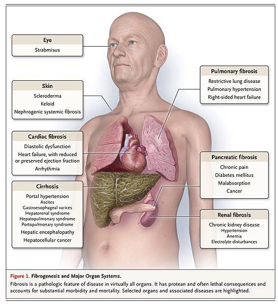
Disease-related injury in any organ triggers a complex cascade of cellular and molecular responses that culminates in tissue fibrosis. Although this fibrogenic response may have adaptive features in the short term, when it progresses over a prolonged period of time, parenchymal scarring and ultimately cellular dysfunction and organ failure ensue (Figure 1
FIGURE 1
Fibrogenesis and Major Organ Systems.).
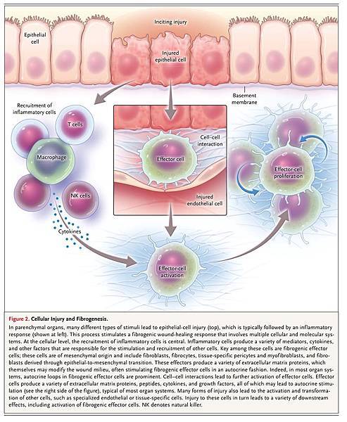
We and others have proposed four major phases of the fibrogenic response (Figure 2
FIGURE 2
Cellular Injury and Fibrogenesis.). First is initiation of the response, driven by primary injury to the organ. The second phase is the activation of effector cells, and the third phase is the elaboration of extracellular matrix, both of which overlap with the fourth phase, during which the dynamic deposition (and insufficient resorption) of extracellular matrix promotes progression to fibrosis and ultimately to end-organ failure.
The fact that diverse diseases in different organ systems are associated with fibrotic changes suggests common pathogenic pathways (Figure 2). This “wounding response” is orchestrated by complex activities within different cells in which specific molecular pathways have emerged. Cellular constituents include inflammatory cells (e.g., macrophages and T cells), epithelial cells, fibrogenic effector cells, endothelial cells, and others. Many different effector cells, including fibroblasts, myofibroblasts, cells derived from bone marrow, fibrocytes, and possibly cells derived from epithelial tissues (epithelial-to-mesenchymal transition) have been identified; there is some controversy regarding the identity of specific effectors in different organs. Beyond the multiple cells essential in the wounding response, core molecular pathways are critical; for example, the transforming growth factor beta (TGF-β) pathway is important in virtually all types of fibrosis.
As fibrosis progresses, myofibroblasts proliferate and sense physical and biochemical stimuli in the local environment by means of integrins and cell-surface molecules; contractile mediators trigger pathological tissue contraction. This chain of events, in turn, causes physical organ deformation, which impairs organ function. Thus, the biology of fibrogenesis is dynamic, although the degree of plasticity appears to vary from organ to organ.
Although we understand many of the cellular and molecular processes underlying fibrosis, there are few effective therapies and fewer that target fibrogenesis specifically. These facts highlight the need for a deeper comprehension of the pathogenesis of fibrogenesis and the translation of this knowledge to novel treatments.
CELLULAR AND MOLECULAR THEMES IN PATHOGENESIS
Acute and chronic inflammation often trigger fibrosis (Figure 2). Inflammation leads to injury of resident epithelial cells and often endothelial cells, resulting in enhanced release of inflammatory mediators, including cytokines, chemokines, and others. This process leads to the recruitment of a wide range of inflammatory cells, including lymphocytes, polymorphonuclear leukocytes, eosinophils, basophils, mast cells, and macrophages. These inflammatory cells elicit the activation of effector cells,1 which drive the fibrogenic process. One example in which an inflammatory lesion drives this cycle is the interstitial nephritis induced by nonsteroidal antiinflammatory drugs (NSAIDs), which culminates in chronic inflammation and the activation of a fibrogenic cascade. In addition, macrophages can play a prominent role in interstitial fibrosis, often driven by the TGF-β pathway.2 However, some inflammatory cells may be protective. For example, certain populations of macrophages phagocytose apoptotic cells that promote the fibrogenic process and activate matrix-degrading metalloproteases.3
Fibroblasts and myofibroblasts have been identified as key fibrosis effectors in many organs, and as such are responsible for the synthesis of extracellular matrix proteins4 (Figure 2). Controversy exists regarding the origin of these cells; for example, myofibroblasts may be derived from fibroblasts or from other mesenchymal cells, such as pericytes.5 Epithelial-to-mesenchymal transition, in which epithelial cells give rise to mesenchymal fibrogenic cells, may play a role. However, the importance of epithelial-to-mesenchymal transition has been actively debated.6-9
The matrix proteins that compose the fibrotic scar, which are highly conserved across tissues, consist predominantly of interstitial collagens (types I and III), cellular fibronectin, basement-membrane proteins such as laminin, and other, less abundant elements. In addition, myofibroblasts, which by definition are cells that express smooth-muscle proteins, including actin (ACTA2), are contractile.10 The contraction of these cells contributes to the distortion of parenchymal architecture, which promotes disease pathogenesis and tissue failure.
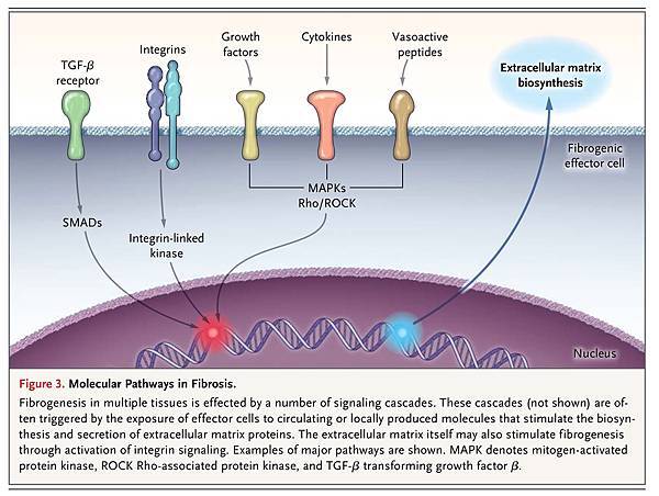
The molecular processes driving fibrosis are wide-ranging and complex (Figure 3
FIGURE 3
Molecular Pathways in Fibrosis.
). The TGF-β cascade, which plays a major role in fibrosis, involves the binding of a ligand to a serine–threonine kinase type II receptor that recruits and phosphorylates a type I receptor. This type I receptor subsequently phosphorylates SMADs, which function as downstream effectors, typically by modulating target gene expression. Although the TGF-β superfamily, which involves multiple signaling cascades, is too complex to review in detail here (see Massagué11), its diversity highlights the complexity of the regulation of fibrosis.
TGF-β is a potent stimulator of the synthesis of extracellular matrix proteins in most fibrogenic cells. TGF-β is synthesized and secreted by inflammatory cells and by effector cells, thereby functioning in both an autocrine and paracrine fashion. The complexity of the TGF-β system is illustrated by its interactions with other cell-signaling pathways.12 For example, TGF-β stimulates sonic hedgehog signaling in lung fibroblasts, and sonic hedgehog signaling, in turn, regulates fibroblast function.13 Another example of the complexity of the TGF-β system emerges from study of the extracellular activation of TGF-β in an inactive complex with a latency-associated peptide, which is subsequently activated by the αvβ6 integrin.14
The molecular systems involved in fibrosis are so expansive as to preclude a detailed discussion (see Figure 3 for an overview of several important molecular pathways). Platelet-derived growth factor (PDGF), connective-tissue growth factor (CTGF), and vasoactive peptide systems (especially angiotensin II and endothelin-1)15 play important roles. Among vasoactive systems, endothelin plays a role in fibrosis in virtually all organ systems, acting through G-protein–coupled endothelin-A or endothelin-B cell-surface receptors or both.16 Furthermore, angiogenic pathways may be important in fibrosis.17 Finally, it is clear that integrins, which link extracellular matrix to cells, are critical in the pathogenesis of fibrosis.18,19
When injury and inflammatory responses are abrogated, resorption of extracellular matrix proteins occurs, promoting organ repair. When chronic injury persists, the unremitting activation of effector cells results in the continuous deposition of extracellular matrix, progressive scarring, and organ damage. Thus, fibrogenesis involves the interplay between factors that promote the biosynthesis, deposition, and degradation of extracellular matrix proteins. Typically, matrix synthesis is counterbalanced by matrix-degrading metalloproteases.3,20,21 In addition, fibrogenesis is governed by pathways that eliminate effectors (e.g., by means of senescence, apoptosis, or autophagy). For example, the apoptosis of hepatic stellate cells is associated with the reversal of fibrosis.22
An understanding of the role of genetics in the pathogenesis of fibrosis is emerging. For instance, in the kidney, fibrosis is a prominent feature of karyomegalic interstitial nephritis, which is caused by mutations in the gene encoding Fanconi anemia-associated nuclease 1 (FAN1).23 In the liver, PNLAP3 is important in fibrosis that is mediated by ethanol and associated with fatty liver disease.24,25 A number of candidate genes may be important in cases of fibrosis that are mediated by infection with hepatitis C virus (HCV).26 Mutations in TERT (c:2768C→T), the gene encoding telomerase reverse transcriptase, and MUC5B, which encodes mucin, are associated with pulmonary fibrosis.27-29
The epigenetic regulation of gene expression, which includes but is not limited to DNA methylation, post-translational modifications of the histone protein constituents of chromatin, and regulatory noncoding RNAs (e.g., microRNAs [miRs]), is important in fibrosis. For example, fibroblasts from patients with idiopathic pulmonary fibrosis have been found to have global changes in DNA methylation, changes that are not seen in fibroblasts from normal lungs.30 The miRs, which play an ever-expanding role in gene regulation, are also involved in the pathogenesis of fibrosis. In diabetic nephropathy, TGF-β promotes the expression of miR-192, which results in collagen deposition,31 and miR-19b regulates TGF-β signaling in hepatic stellate cells.32 In the heart, miR-21, miR-29, miR-30, and miR-133 participate in the remodeling of the myocardial matrix.33
MECHANISMS AND ADVERSE CLINICAL EFFECTS
Cardiac Fibrosis
The heart undergoes extensive structural and functional remodeling in response to injury, central to which is the hypertrophy of cardiac myocytes, with excessive deposition of extracellular matrix.34 Myocardial fibrosis is commonly categorized as one of two types: reactive fibrosis or replacement fibrosis. Reactive fibrosis occurs in perivascular spaces and corresponds to similar fibrogenic responses in other tissues; replacement fibrosis occurs at the site of myocyte loss.
In the heart, fibrosis is attributed to cardiac fibroblasts, the most abundant cell type in the myocardium. These cells are derived from fibroblasts that are native to the myocardium, from circulating fibroblasts, and from fibroblasts that emerge from epithelial-to-mesenchymal transition.35,36 All these cell types proliferate and differentiate into myofibroblasts in response to injury (Figure 2), a process that is driven by classic factors such as TGF-β1, endothelin-1, and angiotensin II.37 Cross-talk and feedback also occur between cells — in this case, between activated fibroblasts and cardiomyocytes — which further fuel fibrogenesis.38
Cardiac fibrosis contributes to both systolic and diastolic dysfunction and to perturbations of electrical excitation; it also disrupts repolarization (Figure 1).39 Proarrhythmic effects are the most prominent. Collagenous septa in the failing heart contribute to arrhythmogenesis by inducing a discontinuous slowing of conduction.40 Areas of arrhythmogenic fibrosis slow conduction through junctions in the heterocellular gap that couple fibroblasts and cardiomyocytes.41 Endocardial breakthrough of microreentrant circuits occurs as a result of the heterogeneous spatial distribution of fibrosis42 and the triggering of activity caused by the depolarization of myocytes by electrically coupled myofibroblasts.43
Fibrotic scarring in the heart correlates strongly with an increased incidence of arrhythmias and sudden cardiac death.44 For example, a 3% increase in the extracellular volume fraction of fibrous tissue (measured by means of magnetic resonance imaging after the administration of gadolinium) is associated with a 50% increase in the risk of adverse cardiac events.45
Hepatic Fibrosis
Hepatic fibrosis typically results from an inflammatory process that affects hepatocytes or biliary cells. Inflammation leads to the activation of effector cells, which results in the deposition of extracellular matrix. Although a variety of effectors synthesize extracellular matrix in the liver, hepatic stellate cells appear to be the primary source of extracellular matrix. Abundant evidence suggests that the stellate cell is pericyte-like, undergoing a transformation into a myofibroblast in response to injury.10
In the liver, multiple cell types, including stellate cells, endothelial cells, Kupffer cells, bile-duct cells, and immune cells, orchestrate the cellular and molecular response to injury.46 Numerous molecular pathways, similar to those found in other organs, are involved. A pathway that appears to be unique to the liver involves toll-like receptor 4 (TLR4)47; TLR4 is activated on the surface of stellate cells by intestinal bacterial lipopolysaccharides derived from the gut (i.e., translocated bacteria), triggering cell activation and fibrogenesis and thereby linking fibrosis to the microbiome.48 TLR4 expression is associated with portal inflammation and fibrosis in patients with fatty liver disease.49
The end result of hepatic fibrogenesis is cirrhosis, an ominous parenchymal lesion that underlies a wide range of devastating complications that have adverse effects on survival (Figure 1). Portal hypertension, a devastating result of injury, develops during the fibrogenic response after disruption of the normal interaction between sinusoidal endothelial cells and hepatic stellate cells; the resulting activation and contraction of pericyte-like stellate cells leads to sinusoidal constriction and increased intrahepatic resistance. This increase in resistance in turn activates abnormal signaling by smooth-muscle cells in mesenteric vessels. An increase in angiogenesis and collateral blood flow follows, resulting in an increase in mesenteric blood flow and a worsening of portal hypertension.50 The major clinical sequelae of portal hypertension, variceal hemorrhage and ascites, emerge relatively late, after the portal pressure rises to a hepatic venous pressure gradient of more than 12 mm Hg.50
Renal Fibrosis
Events that initiate renal fibrosis are diverse, ranging from primary renal injury to systemic diseases.51,52 The kidneys are susceptible to hypertension and diabetes, the two leading causes of renal fibrosis. As is true in other organs, fibrosis of the kidney is mediated by cellular elements (e.g., inflammatory cells) and molecular elements (e.g., cytokines, TGF-β1, CTGF, PDGF, and endothelin-1) (Figure 2 and Figure 3).51-54 The intrarenal renin–angiotensin–aldosterone axis is particularly important in hypertension-induced fibrosis.54
The kidney has a unique cellular architecture that consists of the glomeruli, tubules, interstitium, and capillaries. Injury at any of these sites triggers the deposition of extracellular matrix.37 The location of the initial injury is an important determinant of the clinical consequences. Injuries that initially target glomeruli elicit patterns of disease that are different from those that are elicited by injuries to the tubular–interstitial environment. For example, NSAIDs, urinary obstruction, polycystic kidney disease, and infections can provoke tubulointerstitial fibrosis,43 whereas glomerular immune deposition (e.g., the deposition of IgA) leads to glomerulonephritis.44 Glomeruli and podocytes are sensitive to systemic and local immunologic insults45; high glomerular capillary pressure, exacerbated by systemic hypertension and diabetes, leads to proteinuria, the activation of cytokines and complement, and the infiltration of immune cells, resulting in epithelial cell and interstitial fibrosis.46
Glomerular fibrosis, regardless of the cause, diminishes renal blood flow, which leads to hypoxia and the activation of hypoxia-inducible factor 1, which in turn triggers nephron collapse and fibrotic replacement by means of rarefaction.47 The renal interstitium and capillaries contribute substantially to tubulointerstitial disease, as peritubular pericytes migrate into the interstitium, where they are transformed into myofibroblasts.48
Regardless of the initiating insult, renal fibrosis leads to loss of function and organ failure (Figure 1). Homeostasis can be maintained with a glomerular filtration rate as low as approximately 10% of the normal rate. As the mechanisms maintaining homeostasis are progressively disrupted, anemia develops and the regulation of electrolyte balance and pH is disrupted.
Pulmonary Fibrosis
Pulmonary fibrosis occurs in association with a wide range of diseases, including scleroderma (systemic sclerosis), sarcoidosis, and infection, and as a result of environmental exposures (e.g., silica dust or asbestos), but in most patients it is idiopathic and progressive. Idiopathic pulmonary fibrosis is characterized by progressive fibrosis without substantial inflammation.55 Its pathogenesis appears to be unlike that of other fibrosing diseases and is poorly understood. Injury to alveolar epithelial cells activates pulmonary fibroblasts, provoking their transformation to matrix-producing myofibroblasts.56 Activated lung fibroblasts may cause apoptosis of alveolar cells, which leads to further fibroblast activation and a vicious cycle of injury and effector-cell activation (Figure 2). Research has focused on TGF-β signaling (Figure 3)57 and the interstitial pericytes present in lung fibrosis.58
Pulmonary fibrosis is characterized by parenchymal honeycombing, reduced lung compliance, and restrictive lung function (Figure 1). Fibrosis of the interstitial spaces hinders gas exchange, culminating in abnormal oxygenation and clinical dyspnea. Progressive pulmonary fibrosis also leads to pulmonary hypertension, right-sided heart failure, and ultimately respiratory failure.
Other Forms of Fibrosis
Fibrosis also occurs in the joints, bone marrow, brain, eyes, intestines, peritoneum and retroperitoneum, pancreas, and skin, and in these cases is driven by typical cellular and molecular processes (Figure 2 and Figure 3). Retroperitoneal fibrosis is a rare condition characterized by inflammation and fibrosis in the retroperitoneal space; most cases are idiopathic, but secondary causes include drugs, infections, autoimmune and inflammatory stimuli, and radiation. Patients may present with pain, and the major clinical sequelae of this condition are related to its involvement with structures in the retroperitoneum, including arteries (leading to acute and chronic renal failure) and ureters (leading to hydronephrosis). Currently, treatment of this primary fibrosing disorder is not available. In certain cancers, fibrosis is linked to TGF-β-integrin signaling.59 In scleroderma, the prototypical fibrosing skin disease, skin fibroblasts and myofibroblasts are activated through the TGF-β–SMAD signaling pathway.60 Nephrogenic systemic fibrosis, a debilitating condition that is marked by widespread organ fibrosis, occurs in patients with renal insufficiency who have been exposed to gadolinium-based contrast material. Initial systemic inflammatory-response reactions and the reaction of gadolinium (Gd3+) ions with circulating proteins and heavy metals lead to the deposition of insoluble elements in tissue.61 Since no effective therapies have been identified, prevention is key.61 A recently recognized IgG4-related disease appears to involve autoimmune-driven inflammation that provokes fibrosis in multiple organs, including the pancreas, retroperitoneum, lung, kidney, liver, and aorta.62
THERAPY
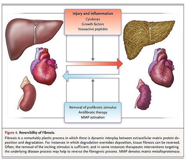
Fibrosis and resultant organ failure account for at least one third of deaths worldwide.63 Since fibrosis is common and has adverse effects in all organs, it is an attractive therapeutic target. Contrary to the widely held perception that scar tissue is permanent, the available evidence points to the highly plastic nature of organ fibrosis; it is not irreversible “scar” but an actively remodeled tissue component that can, under certain circumstances, regress. Fibrosis occurs by means of a dynamic process that involves the synthesis and deposition of extracellular matrix, and its reversion occurs by means of the elimination of effector cells and shifts in the balance of matrix synthesis and degradation (Figure 4
FIGURE 4
Reversibility of Fibrosis.
). Although it is not clear what pathogenic or clinical factors promote reversibility, the regression of fibrosis has been shown to lead to improved clinical outcomes. Elimination of the inciting stimulus is the first and most efficacious approach.
Fibrosis of parenchymal tissue usually progresses slowly, which suggests that therapy may be required for extended periods; slowing the progression of fibrosis may be a more realistic therapeutic goal than eliminating it. One of the challenges in assessing the therapeutic response is that there are few noninvasive means of measuring fibrosis (e.g., blood or imaging tests; biopsy is typically the most reliable approach) or monitoring the effects of therapeutic intervention.
The best indication that fibrosis is reversible and that this reversibility has positive effects on clinical outcomes is based on the treatment of liver disease64; in patients with cirrhosis who are infected with hepatitis B virus (HBV), antiviral therapy reduces fibrosis, reverses cirrhosis,65 and reduces the incidence of clinical complications.66 Pirfenidone has been shown to slow the reduction in forced vital capacity and reduce mortality, raising the possibility of a reversal in fibrosis.
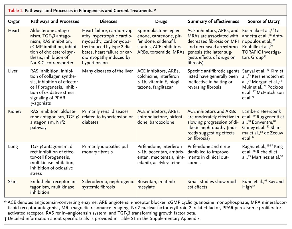
Often, promising preclinical studies are not borne out in clinical trials in terms of both expected efficacy and unexpected side effects. Thus, at the current time, specific antifibrotic therapies are limited (see Table 1
TABLE 1
Pathways and Processes in Fibrogenesis and Current Treatments.
, which summarizes core concepts from large, completed clinical trials targeting fibrosis in humans (including both positive and negative results), and Table S1 in the Supplementary Appendix, which includes more detailed information about completed clinical trials and is available with the full text of this article at NEJM.org). Several agents targeting specific fibrotic pathways in the liver and the lung have been examined, placing the studies of the liver and lung ahead of studies of other organ systems in terms of the goal to specifically confront fibrosis. Novel approaches to the treatment of fibrosis that are based on an extensive body of preclinical data are anticipated in the coming years (see Table S2, which highlights early phase trials that target less well established, although potentially important, pathways in fibrosis).
Heart
Pharmacologic therapies in clinical use for heart failure that target the primary underlying disease appear to have a secondary effect on fibrosis. Examples include angiotensin-converting–enzyme (ACE) inhibitors, statins, aldosterone antagonists, and emerging therapies, such as histone deacetylase inhibitors. Such therapies, which are known to promote beneficial “reverse remodeling,” ameliorate fibrosis, reduce the burden of ventricular arrhythmia, slow the rate of ventricular tachycardias,93,94 and reduce the incidence of sudden death.39
A promising idea for the treatment of cardiac fibrosis is based on the premise that cardiac fibroblasts can be reprogrammed into cardiomyocyte-like cells95,96 (Table S2 in the Supplementary Appendix) that would promote normal tissue regeneration; in a murine model of myocardial infarction and fibrosis, the retroviral expression of specific transcription factors in the myocardium reprogrammed cells that acquired spontaneous contractile and electrophysiological properties resembling those of cardiomyocytes, leading to global improvements in contractile function. It is not yet known whether this type of therapy can be used in humans.
Kidney
Like the therapies used to treat cardiac fibrosis, those typically used to prevent renal fibrosis target the underlying disease processes and as such involve the treatment of hypertension and diabetes. One target is the renin–angiotensin system. This approach involves the use of ACE inhibitors and angiotensin-receptor blockers that ameliorate renal damage and fibrosis through multiple pathways, including the suppression of the actions of TGF-β.79 Therapies based on the antagonism of aldosterone that make use of mineralocorticoid receptor antagonists have been shown to inhibit or slow the progression of fibrosis in humans.97 Novel approaches to the treatment of fibrosis of the kidneys include those that target bone morphogenetic protein-7, NADPH oxidase (NOX) (NOX1 and NOX4), and the SMAD3 and SMAD4 pathways (Table S2 in the Supplementary Appendix).98
Liver
The process of hepatic fibrosis is dynamic. Since hepatocytes are capable of regeneration, liver fibrosis may be especially amenable to therapeutic intervention, and even cirrhosis can be reversed.66,99-101 Eradication of HCV infection, antiviral therapy for HBV infection, glucocorticoid therapy for autoimmune hepatitis, phlebotomy for hemochromatosis, relief of biliary obstruction, and cessation of alcohol consumption in alcoholic hepatitis each clearly reverses fibrotic change, and many of these treatments improve clinical outcomes.66,99,100,102
A number of potential antifibrotic therapies targeting specific pathways have been studied in human liver disease (Table S2 in the Supplementary Appendix). For example, colchicine suppresses collagen secretion and theoretically prevents fibrosis.46 Interferon γ-1b and the peroxisome proliferator-activated receptor γ ligand, farglitazar, which inhibit stellate cell-mediated fibrogenesis, were studied in patients infected with HCV that was unresponsive to primary antiviral therapy, but no beneficial effects on fibrosis were noted.46 Other agents, including polyenephosphatidyl choline, silymarin, and ursodeoxycholic acid, have similarly shown no benefit.46 Vitamin E had modest effects on histologic fibrosis in patients with nonalcoholic steatohepatitis.72
Lung
The lung presents special challenges with regard to therapy targeting fibrosis. On the one hand, the lung has easily measured clinical features that allow for assessment of lung function, a surrogate for fibrosis. On the other hand, pulmonary fibrosis appears to be less dynamic than fibrosis occurring in other organ systems. Multiple therapies have been tested for pulmonary fibrosis, especially for idiopathic pulmonary fibrosis. Interferon γ-1b showed efficacy in preclinical studies but showed no benefit in human clinical trials.85 Endothelin-receptor antagonists have also showed no benefit. Pirfenidone, a pyridone derivative with antiinflammatory and antifibrotic effects that is available in oral form, reduced disease progression and increased survival in patients with idiopathic pulmonary fibrosis. Its mechanism of action is not completely understood, but it presumably has effects on TGF-β production.88 Nintedanib, a multikinase inhibitor, slowed disease progression in a similar cohort.89 It is important to note that the outcomes in these trials were based on clinical results; reductions in lung fibrosis per se have not been definitively demonstrated.
FUTURE DIRECTIONS
Fibrosis is a hallmark of pathologic remodeling in numerous tissues and a contributor to clinical disease. There is a great deal of interest in identifying means of slowing, arresting, or even reversing the progression of tissue fibrogenesis. Thus, it is important to understand the central mechanisms underlying the fibrogenic process. Common themes implicating conserved cellular and molecular pathways have emerged. A major conserved cellular element is the activated fibroblast, also known as a myofibroblast, which produces abundant amounts of extracellular matrix. Some of the major conserved molecular processes involve TGF-β, PDGF, CTGF, vasoactive compounds (endothelin-1 and angiotensin II), and integrin–extracellular matrix signaling pathways. The fact that tissue fibrosis is remarkably plastic suggests that many of these major elements of disease pathogenesis may emerge as targets of novel therapeutic interventions.




 留言列表
留言列表
 線上藥物查詢