儘管限制飲食,慢性口服carnitine依然會升高TMAO
Abstract
Recent studies have implicated trimethylamine N-oxide (TMAO) in atherosclerosis, raising concern about l-carnitine, a common supplement for patients with inborn errors of metabolism (IEMs) and a TMAO precursor metabolized, in part, by intestinal microbes. Dietary meat restriction attenuates carnitine-to-TMAO conversion, suggesting that TMAO production may not occur in meat-restricted individuals taking supplemental l-carnitine, but this has not been tested. Here, we mine a metabolomic dataset to assess TMAO levels in patients with diverse IEMs, including organic acidemias. These data were correlated with clinical information and confirmed using a quantitative TMAO assay. Marked plasma TMAO elevations were detected in patients treated with supplemental l-carnitine, including those on a meat-free diet. On average, patients with an organic acidemia had ~45-fold elevated [TMAO], as compared to the reference population. This effect was mitigated by metronidazole therapy lasting 7 days each month. Collectively, our data show that TMAO production occurs at high levels in patients with IEMs receiving oral l-carnitine. Further studies are needed to determine the long-term safety and efficacy of chronic oral l-carnitine supplementation and whether suppression or circumvention of intestinal bacteria may improve l-carnitine therapy.
Introduction
Supplemental l-carnitine is widely used in the treatment of inborn errors of metabolism (IEMs). Daily oral doses of high levels (~100 mg l-carnitine/kg body weight) of this compound are routinely employed in the treatment of organic acidemias and primary carnitine uptake deficiency (OMIM212140) and less frequently in the treatment of multiple other IEMs including, but not limited to, urea cycle defects, fatty acid oxidation disorders, mitochondrial disease, maple syrup urine disease (OMIM248600), and lysinuric protein intolerance (OMIM222700) (Mori et al. 1990; Davies et al. 1991; Kolker et al. 2011; Sebastio et al. 2011; Magoulas and El-Hattab 2012; Mescka et al. 2015; Tischner and Wenz 2015). Carnitine plays an essential role in mitochondrial shuttling; supplementation is therefore aimed at replenishing depleted carnitine stores and conjugating and removing the toxic buildup of organic acids. No long-term placebo-controlled studies have been completed to test the safety and efficacy of this therapy, but for many IEMs there are strong theoretical and observational data advocating for the use of l-carnitine supplementation (Walter 2003; Winter 2003; Nasser et al. 2012). In individuals without an IEM, carnitine is naturally supplied primarily through consumption of meat, with the remainder produced by an endogenous synthesis pathway (Rebouche 2004).
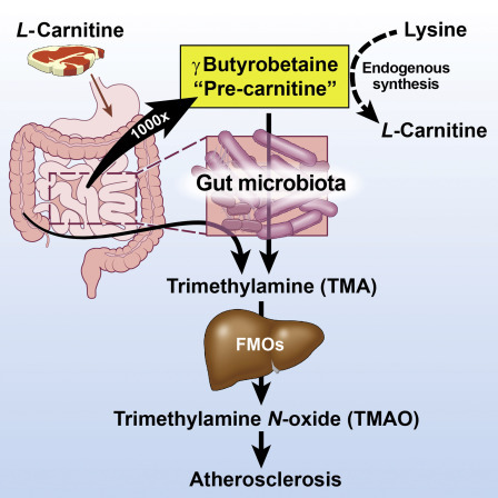
When orally consumed, l-carnitine can be metabolized by intestinal bacteria to form trimethylamine (TMA), a volatile compound associated with a pungent fishy odor (Zhang et al. 1999). TMA is readily absorbed in the gastrointestinal tract and further processed by the hepatic enzyme flavin monooxygenase 3 (FMO3) to produce trimethylamine N-oxide (TMAO) (Lang et al. 1998) (Fig. 1a). Carnitine to TMAO conversion varies widely between individuals with reports that, for some, as much as ~50% of ingested carnitine is eventually excreted in the urine as TMAO (Rebouche 1991). Interindividual differences in the intestinal microbiome may explain some of the variation in conversion rates. Individuals treated with antibiotics targeting intestinal bacteria or those that adhere to a vegan or vegetarian diet have significantly lower baseline levels of plasma TMAO and do not produce appreciable levels of this compound when given oral l-carnitine for short periods of time (Koeth et al. 2013).
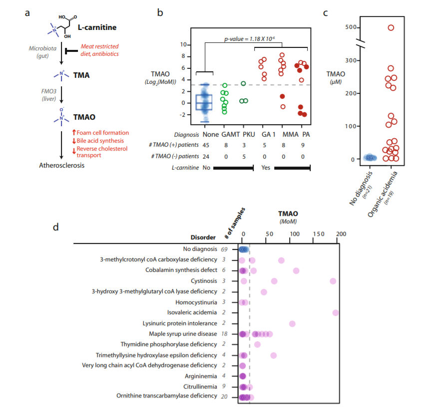
High levels of TMAO detected in the plasma of patients with IEMs requiring l-carnitine supplementation. (a) The diagram illustrates the current model for l-carnitine microbial metabolism and its association with atherosclerosis (Modified from (Backhed 2013)). (b) Plasma TMAO levels discovered via metabolomic analysis are plotted in terms of log2-transformed multiples of the median (MoM) of the reference population. Samples from patients without a diagnosis are plotted with blue dots (n = 69), and the gray dashed lineindicates the highest TMAO value from this group. Filled circles indicate the organic acidemia patients treated with antibiotics within the 30 days prior to sampling. GAMT guanidinoacetate methyltransferase deficiency, PKU phenylketonuria, GA 1 glutaric acidemia type 1, MMA methylmalonic acidemia, PA propionic acidemia. Below the plot is listed the number of patients for which TMAO was confidently identified (TMAO(+)) or below the level of detection (TMAO(−)) for the metabolomic platform. (c) A subset of specimens was reanalyzed using a quantitative TMAO analysis. Organic acidemia includes MMA, PA, and GA 1 patients. (d) The TMAO values from metabolomic analyses of plasma samples from additional IEM patients are shown. The gray dashed line indicates the highest TMAO level seen in the reference population (“no diagnosis”)
Recent studies have identified plasma TMAO as a dose-dependent risk factor for cardiovascular disease that may advance atherosclerosis through the promotion of cholesterol storage in macrophages (Wang et al. 2011; Koeth et al. 2013; Tang et al. 2013). These findings hold obvious implications about the long-term consequences of chronic oral l-carnitine supplementation in healthy individuals on a standard western diet. However, in patients with IEMs, carnitine-TMAO conversion is not established, and it has been postulated that dietary protein restriction may mitigate gut microbial breakdown of supplemental l-carnitine, similar to what is seen in vegan/vegetarian individuals. Additionally, for the organic acidemias, methylmalonic and propionic acidemia (OMIM 251000 and 606054, respectively), monthly prophylactic treatment with the antibiotic metronidazole is also commonly used to reduce intestinal bacteria and the affiliated production of propionate, and this may confer the unexpected benefit of reducing TMAO production as well (Thompson et al. 1990). Taken together, the outcome of carnitine supplementation is not obvious for many IEMs, especially the organic acidemias, and to date, no studies have assessed the levels of TMAO in this population.
To test the hypothesis that chronic oral supplemental l-carnitine could lead to TMAO production in patients undergoing standard treatment for an organic acidemia, we mined a metabolomic dataset previously generated by our lab, which contains metabolic information on patients with a wide variety of IEMs (Miller et al. 2015). Findings from this analysis were correlated with clinical data and confirmed using a targeted tandem mass spectrometry (MS/MS) assay that allowed absolute quantification of TMAO.
Methods
The methods and a general overview of findings from the metabolomic analyses in this report have been previously described (Miller et al. 2015). Briefly, an untargeted MS/MS-based metabolomic platform was used to semiquantitatively analyze ~900 unique small molecule plasma analytes in each of 190 patient specimens including 69 patients without a diagnosed genetic disorder. This latter patient group is likely comprised of individuals on a standard omnivorous western diet and not on supplemental carnitine; we refer to this as the “reference population” for the remainder of the manuscript. Raw spectral intensity values were normalized to allow comparison across independent MS/MS batches, and these normalized values were then used to scale all analyte values to the median of the reference population (n = 69). The median age of the reference population was 5 years (inner quartile range (IQR) = 2.4–13.1). A two-tailed heteroscedastic student’s t-test was used to compare log2-transformed data. Linear regression analysis was completed using the freely available statistical package R.
For quantitative TMAO analysis, plasma specimens were diluted 1:10 in acetonitrile containing a spiked isotopic standard, TMAO-D9 (Cambridge isotope DLM-4779-1). Specimens were clarified by centrifugation and supernatants were subjected to liquid chromatography tandem mass spectrometry (LC-MS). Chromatographic separation was achieved using an Aquity UPLC (Waters) equipped with an Atlantis HILIC Silica 5 μm 1 × 100 mm column (Waters) and an isocratic mobile phase—80% acetonitrile 0.1% formate and 20% 50 mM ammonium acetate pH 4.8. MS analysis was completed in positive ion mode using an Aquity TQ tandem MS (Waters) with the following transitions: TMAO 76.1 > 58.1 m/z and TMAO-D9 85.3 > 66.2 m/z. Quantitative values were calculated by fitting a sample’s TMAO/TMAO-D9 response factor to a linear model generated via the analysis of six standards ranging in concentration from 0.2 to 625 μM TMAO.
Results
In a prior study, we explored the clinical utility of an untargeted metabolomic platform in the retrospective analysis of plasma specimens collected from patients with a wide range of IEMs, as well as individuals without a biochemical diagnosis (Miller et al. 2015). This sample set included specimens from multiple unrelated patients receiving treatment for organic acidemias, including glutaric acidemia type 1 (OMIM#231670), methylmalonic acidemia, and propionic acidemia. This group represents the cohort of individuals in our sample set most likely to be on l-carnitine supplementation while also adhering to a meat-restricted diet. Clinical information was available for the majority of the subjects, and for these cases we confirmed daily oral l-carnitine supplementation and restrictions of dietary meat consumption (Table 1).
Table 1
Clinical information for patients in the organic acidemia cohort
| ID | Disordera | Age at sampling (years) | Daily l-carnitine dosage (mg/kg body weight)b | Recent antibiotics exposurec | TMAOd | 3-MHd |
|---|---|---|---|---|---|---|
| PAT1 | GA 1 | 9.8 | Unknown | Unknown | 18.14 | No ID |
| PAT2 | GA 1 | 3.6 | 88 | No | 31.14 | No ID |
| PAT3 | GA 1 | 4.8 | 91 | No | 73.48 | No ID |
| PAT4 | GA 1 | 6.3 | 75 | No | 182.96 | No ID |
| PAT5 | GA1 | 3.6 | Unknown | Unknown | 142.01 | No ID |
| PAT6 | MMA | 10.6 | 83 IV | Vanc./Zosyn | 2.17 | 0.97 |
| PAT7a | MMA | 4.9 | 100 | No | 117.30 | No ID |
| PAT7b | MMA | 5.0 | 100 | No | 32.83 | No ID |
| PAT8 | MMA | 2.1 | 103 | No | 0.64 | No ID |
| PAT9 | MMA | 6.1 | Unknown | Unknown | 78.09 | No ID |
| PAT10a | MMA | 36.3 | 19 | No | 312.83 | 3.22 |
| PAT10b | MMA | 36.7 | 19 | No | 164.24 | 19.13 |
| PAT11 | MMA | 8.7 | 97 | No | 50.35 | 0.96 |
| PAT12 | PA | 0.9 | 109 | Metron. (7 days/month) | 0.25 | 0.19 |
| PAT13 | PA | 2.1 | 51 | Metron. (7 days/month) | 0.62 | No ID |
| PAT14 | PA | 3.9 | 145 | Metron. (7 days/month) | 0.28 | No ID |
| PAT15 | PA | 2.0 | 96 | Augmen. | 50.13 | 0.66 |
| PAT16a | PA | 4.5 | Unknown | Unknown | 113.43 | No ID |
| PAT16b | PA | 4.9 | Unknown | Unknown | 127.24 | 0.27 |
| PAT17 | PA | 0.4 | 80 | Metron. (5 days/month) | 73.26 | 0.19 |
| PAT18 | PA | 3.4 | 75 | Metron. (5 days/month) | 85.45 | No ID |
| PAT19 | PA | 7.4 | 84 | No | 15.25 | 0.78 |
a GA1 glutaric acidemia type 1, MMA methylmalonic acidemia, PA propionic acidemia
bIV indicates a patient whom was taken off oral carnitine (prior chronic PO dose is listed) and instead was on intravenous carnitine replacement for 10 days prior to sampling as a result of illness and inpatient hospitalization
cAntibiotic treatments within 30 days of sampling are listed. Metron. = oral metronidazole; Vanc./Zosyn = intravenous Vancomycin and Zosyn.; Augmen. = Augmentin, 10-day course. Treatment regimens are listed in parenthesis, so “7 days/month” = antibiotic is given for 7 days out of the month
dValues are reported as multiples of the median value of the analyte determined in the reference population. Compounds below the level of detection = “No ID”; 3-MH = 3-methylhistidine
Dietary compliance was independently audited by further mining of the metabolomic data. 3-methylhistidine (3-MH) is a muscle breakdown product that is significantly elevated following meat consumption (Cross et al. 2011). This compound was positively identified in the plasma of 77% of the patients in our reference population, whereas for most organic acidemia patients, 3-MH was undetectable (73% of patients) or well below the median value for the reference population, thus suggesting compliance with a meat-restricted diet for the majority of subjects (Table 1).
Despite the apparent compliance with meat restriction, metabolomic analysis identified marked elevations of TMAO in the majority of patients with an organic acidemia (Fig. 1b). For 14 of 19 patients with an organic acidemia, TMAO levels were greater than 15 times the median level of the reference population (multiples of the median, MoM) with an overall average TMAO level in the organic acidemia cohort of 65 MoM (p-value = 2.19 × 10⁻⁸). Metabolomic findings were confirmed by reanalysis of a subset of specimens using a targeted MS/MS assay that provided absolute quantification and lower limits of detection (Fig. 1c). Importantly, quantitative values generated by this method correlated with metabolomic data (R 2 = 0.68) and confirmed the significant elevations of TMAO in many organic acidemia patients. Patients with an organic acidemia had an average plasma TMAO concentration of 120 μM (IQR = 24.3–194.6 μM) as compared to 2.75 μM (IQR = 1.1–4.5 μM) in the reference population. The average TMAO concentration detected in our reference population was similar to previous reports of baseline TMAO levels in healthy individuals on an omnivorous diet and not taking an l-carnitine supplement (TMAO = ~3–4 μM) (Koeth et al. 2013).
More sporadic patterns of extreme TMAO elevations were noted in additional IEM patient groups, with the pattern likely reflecting the sporadic use of l-carnitine supplementation in the treatment of these diseases. Examples of patients with extreme plasma TMAO levels include those with cobalamin synthesis disorders, cystinosis (OMIM219800), isovaleric acidemia (OMIM243500), and maple syrup urine disease (OMIM248600) (Fig. 1d). It is notable that multiple patients in our dataset were not on supplemental l-carnitine and consequently had very low levels of plasma TMAO; these include the patients in the reference population who did not have a diagnosis (n = 69) or patients with IEMs that are not typically treated with l-carnitine supplementation (e.g., guanidinoacetate methyltransferase deficiency (OMIM612736; n = 8) and phenylketonuria (OMIM261600; n = 8)). For nearly all patients within this group, TMAO was either below the level of detection or detected at a low-to-moderate level (Fig. 1b).
Given the established relationship between TMAO production and intestinal microbiota, we were interested in the effects of antibiotics in our organic acidemia cohort. Overall, seven patients were prescribed antibiotics, typically metronidazole, at the time of sampling or within the 30 days prior to sampling (filled dots in Fig. 1b and Table 1). Challenging this analysis was our inability to confirm compliance or timing of metronidazole treatments relative to sampling. However, it is noteworthy that within the organic acidemia cohort, five patients had biochemical evidence of attenuated microbial carnitine-TMAO conversion, e.g., low-to-normal plasma TMAO levels. Of these five, four were on antibiotic treatments immediately preceding or during sampling (PAT12-14 and PAT6; Table 1). Patients on 7 days per month metronidazole therapy had normalized TMAO levels (PAT12-14), whereas two patients on 5 days per month metronidazole therapy had extreme TMAO elevations (PAT 17-18) suggesting that the duration of antibiotic therapy may impact microbial l-carnitine degradation (Table 1).
Discussion
The identification of high levels of TMAO in patients with an organic acidemia is clinically important for two reasons. First, it disproves the hypothesis that dietary protein/meat restriction may protect these patients from intestinal microbial conversion of carnitine to TMAO. The TMAO levels in most patients with an organic acidemia were well above the levels observed in even the most extreme examples in our reference population. Given that improved therapies have increased the survival of individuals with organic acidemias, these data indicate that long-term studies of oral carnitine supplementation in this population are needed to understand the risk of atherosclerosis. Second, our findings also have important implications regarding the efficacy of carnitine therapy in patients with IEMs. The bioavailability of supplemental l-carnitine has been previously noted to be <25%, and trials replacing oral l-carnitine with venous boluses of l-carnitine have shown promising preliminary results (Winter 2003; Rebouche 2004). Taken together with our findings, gut microbial carnitine-TMAO conversion may be an underappreciated detriment to carnitine therapy. Future improvements may be achieved by cyclic oral antibiotics that limit the microbial conversion of carnitine to TMAO or through the use of parenteral supplementation that would bypass the intestinal microbiome.
In conclusion, these findings demonstrate that oral l-carnitine could be conferring an increased risk to develop atherosclerosis over years of therapy due to chronic and extreme elevations in TMAO. Further investigation of the intestinal microbiome metabolism of orally supplemented l-carnitine in patients with IEMs is needed to answer the following questions raised by this study: (1) Does chronic oral l-carnitine supplementation increase the risk of atherosclerosis in individuals with organic acidemias? (2) Can cyclic administration of an oral antibiotic, such as metronidazole 7 days per month, reduce TMAO levels while increasing plasma carnitine levels in individuals with organic acidemias on chronic oral l-carnitine supplementation? (3) Can periodic parenteral l-carnitine supplementation improve plasma carnitine levels and outcome measures while leading to normal TMAO levels? Due to the pervasive use of oral l-carnitine supplementation in the field of inherited metabolic disease, it is critical that these questions are answered to improve outcomes and minimize the risks of long-term problems in individuals with IEMs.
References
- Backhed F. Meat-metabolizing bacteria in atherosclerosis. Nat Med. 2013;19(5):533–534. doi: 10.1038/nm.3178. [PubMed] [CrossRef]
- Cross AJ, Major JM, Sinha R. Urinary biomarkers of meat consumption. Cancer Epidemiol Biomarkers Prev. 2011;20(6):1107–1111. doi: 10.1158/1055-9965.EPI-11-0048. [PMC free article] [PubMed] [CrossRef]
- Davies SE, Iles RA, Stacey TE, de Sousa C, Chalmers RA. Carnitine therapy and metabolism in the disorders of propionyl-CoA metabolism studied using 1H-NMR spectroscopy. Clin Chim Acta. 1991;204(1–3):263–277. doi: 10.1016/0009-8981(91)90237-7. [PubMed] [CrossRef]
- Koeth RA, Wang Z, Levison BS, et al. Intestinal microbiota metabolism of L-carnitine, a nutrient in red meat, promotes atherosclerosis. Nat Med. 2013;19(5):576–585. doi: 10.1038/nm.3145.[PMC free article] [PubMed] [CrossRef]
- Kolker S, Christensen E, Leonard JV, et al. Diagnosis and management of glutaric aciduria type I--revised recommendations. J Inherit Metab Dis. 2011;34(3):677–694. doi: 10.1007/s10545-011-9289-5.[PMC free article] [PubMed] [CrossRef]
- Lang DH, Yeung CK, Peter RM, et al. Isoform specificity of trimethylamine N-oxygenation by human flavin-containing monooxygenase (FMO) and P450 enzymes: selective catalysis by FMO3. Biochem Pharmacol. 1998;56(8):1005–1012. doi: 10.1016/S0006-2952(98)00218-4. [PubMed] [CrossRef]
- Magoulas PL, El-Hattab AW. Systemic primary carnitine deficiency: an overview of clinical manifestations, diagnosis, and management. Orphanet J Rare Dis. 2012;7:68. doi: 10.1186/1750-1172-7-68. [PMC free article] [PubMed] [CrossRef]
- Mescka CP, Guerreiro G, Hammerschmidt T, et al. L-Carnitine supplementation decreases DNA damage in treated MSUD patients. Mutat Res. 2015;775:43–47. doi: 10.1016/j.mrfmmm.2015.03.008. [PubMed] [CrossRef]
- Miller MJ, Kennedy AD, Eckhart AD, et al. Untargeted metabolomic analysis for the clinical screening of inborn errors of metabolism. J Inherit Metab Dis. 2015;38(6):1029–1039. doi: 10.1007/s10545-015-9843-7. [PMC free article] [PubMed] [CrossRef]
- Mori T, Tsuchiyama A, Nagai K, Nagao M, Oyanagi K, Tsugawa S. A case of carbamylphosphate synthetase-I deficiency associated with secondary carnitine deficiency--L-carnitine treatment of CPS-I deficiency. Eur J Pediatr. 1990;149(4):272–274. doi: 10.1007/BF02106292. [PubMed] [CrossRef]
- Nasser M, Javaheri H, Fedorowicz Z, Noorani Z. Carnitine supplementation for inborn errors of metabolism. Cochrane Database Syst Rev. 2012;2:CD006659. [PubMed]
- Rebouche CJ. Quantitative estimation of absorption and degradation of a carnitine supplement by human adults. Metabolism. 1991;40(12):1305–1310. doi: 10.1016/0026-0495(91)90033-S. [PubMed] [CrossRef]
- Rebouche CJ. Kinetics, pharmacokinetics, and regulation of L-carnitine and acetyl-L-carnitine metabolism. Ann N Y Acad Sci. 2004;1033:30–41. doi: 10.1196/annals.1320.003. [PubMed] [CrossRef]
- Sebastio G, Sperandeo MP, Andria G. Lysinuric protein intolerance: reviewing concepts on a multisystem disease. Am J Med Genet C Semin Med Genet. 2011;157C(1):54–62. doi: 10.1002/ajmg.c.30287.[PubMed] [CrossRef]
- Tang WH, Wang Z, Levison BS, et al. Intestinal microbial metabolism of phosphatidylcholine and cardiovascular risk. N Engl J Med. 2013;368(17):1575–1584. doi: 10.1056/NEJMoa1109400.[PMC free article] [PubMed] [CrossRef]
- Thompson GN, Chalmers RA, Walter JH, et al. The use of metronidazole in management of methylmalonic and propionic acidaemias. Eur J Pediatr. 1990;149(11):792–796. doi: 10.1007/BF01957284. [PubMed] [CrossRef]
- Tischner C, Wenz T. Keep the fire burning: current avenues in the quest of treating mitochondrial disorders. Mitochondrion. 2015;24:32–49. doi: 10.1016/j.mito.2015.06.002. [PubMed] [CrossRef]
- Walter JH. L-carnitine in inborn errors of metabolism: what is the evidence? J Inherit Metab Dis. 2003;26(2–3):181–188. doi: 10.1023/A:1024485117095. [PubMed] [CrossRef]
- Wang Z, Klipfell E, Bennett BJ, et al. Gut flora metabolism of phosphatidylcholine promotes cardiovascular disease. Nature. 2011;472(7341):57–63. doi: 10.1038/nature09922. [PMC free article][PubMed] [CrossRef]
- Winter SC. Treatment of carnitine deficiency. J Inherit Metab Dis. 2003;26(2–3):171–180. doi: 10.1023/A:1024433100257. [PubMed] [CrossRef]
- Zhang AQ, Mitchell SC, Smith RL. Dietary precursors of trimethylamine in man: a pilot study. Food Chem Toxicol. 1999;37(5):515–520. doi: 10.1016/S0278-6915(99)00028-9. [PubMed] [CrossRef]


 留言列表
留言列表
 線上藥物查詢